In one line
Pelvic inflammatory disease is ascending polymicrobial infection of the upper genital tract whose diagnosis is clinical and deliberately over-sensitive, because the cost of a missed case is measured in tubes; treat empirically on minimal criteria with broad-spectrum cover that includes anaerobes, and escalate to admission and drainage when a tubo-ovarian abscess declares itself.
Mechanism & pathophysiology
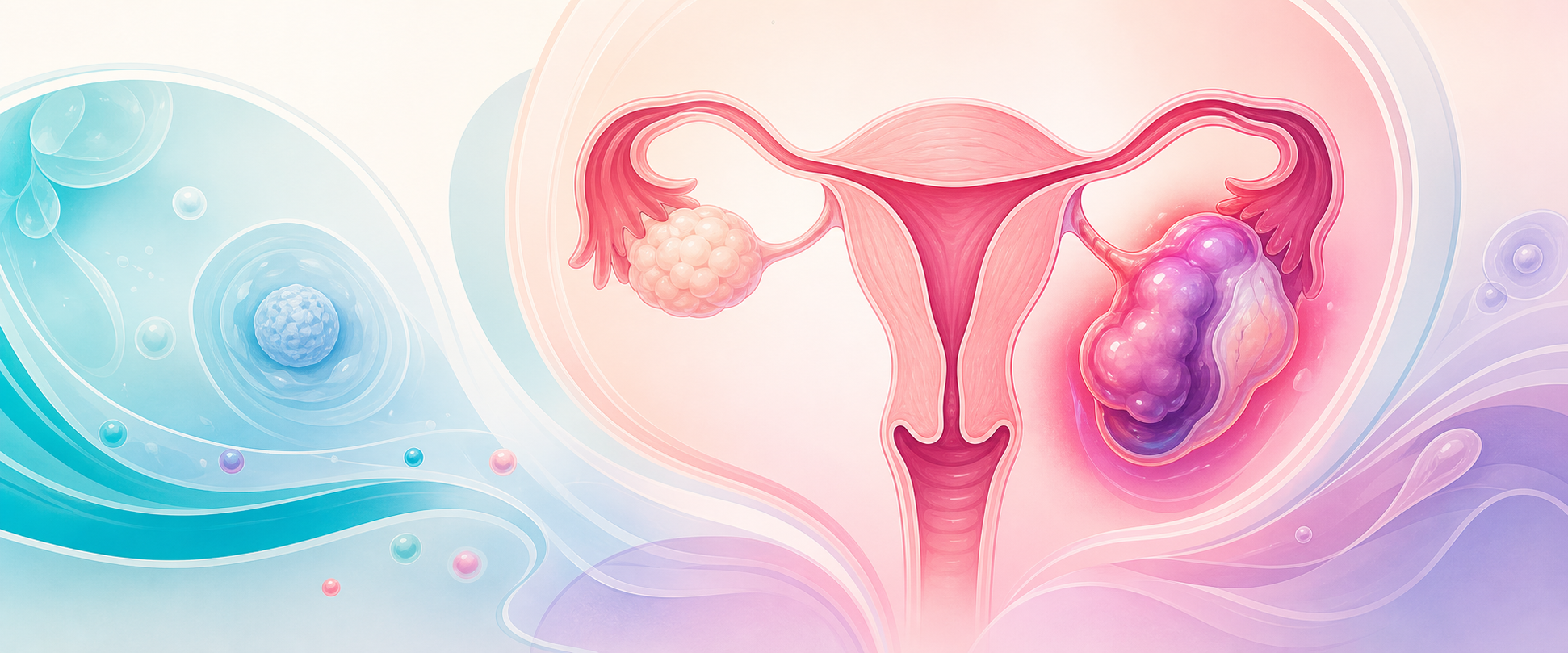
The disease begins at the endocervix and travels upward. Organisms breach the cervical barrier — often around menses, instrumentation or recent intrauterine-device insertion, when the protective mucus plug and the local immunity are disturbed — and ascend the endometrial cavity to the tubes, ovaries and peritoneum. The named stations of that journey are the diagnoses: endometritis, salpingitis, oophoritis, parametritis, and pelvic peritonitis, with a tubo-ovarian abscess as the end-stage walled-off collection.
It is polymicrobial, and that fact dictates the antibiotics. The classical sexually transmitted initiators are Neisseria gonorrhoeae and Chlamydia trachomatis; chlamydia is the single commonest identified cause, found in roughly 14–35% of cases, and in South Africa, where gonococcal prevalence is high, N. gonorrhoeae is a more frequent contributor than in low-prevalence European settings. But the cervical breach is followed by an ascent of the vaginal flora, so anaerobes (Prevotella, Atopobium, Bacteroides), Gardnerella vaginalis and other bacterial-vaginosis-associated organisms colonise the upper tract alongside — which is why a regimen that covers only the gonococcus and chlamydia under-treats. Mycoplasma genitalium is now recognised as a genuine cause of upper-tract infection rather than a bystander, and a substantial proportion of PID is pathogen-negative on lower-tract testing, meaning a negative swab never excludes the disease.
The damage is immunopathological. The host inflammatory response to chlamydia in particular — the cell-mediated reaction to persistent infection — scars the tubal mucosa, flattens the cilia and obliterates the lumen. The mucosal cilia are not merely passengers: their loss is what converts a tube from a transport organ into a blind pouch, so even a tube that remains anatomically patent on a hysterosalpingogram can be functionally useless for ovum pickup. That scarring is the substrate of every long-term sequela: tubal-factor infertility, ectopic pregnancy (a damaged but patent tube traps the conceptus), chronic pelvic pain from dense pelvic adhesions, and recurrent PID, because a once-injured tube defends itself less well against the next ascent. The injury accumulates with each episode and with severity, which is the mechanistic reason early, adequate treatment matters and why repeat infection is so much worse than a single bout — the tubal damage is broadly stepwise with successive episodes.
The microbiology shifts as the disease progresses, and this drives the antibiotic logic over time. The initiating event is frequently the sexually transmitted organism breaching the cervix, but once the upper tract is inflamed and the tissue planes are disrupted, the established and abscess-forming infection becomes increasingly anaerobic — a tubo-ovarian abscess yields a luxuriant mixed anaerobic flora regardless of whether gonorrhoea or chlamydia opened the door. This is why a regimen that covers only the initiating STI is adequate for the earliest endometritis but progressively inadequate as salpingitis matures into an abscess, and why anaerobic cover becomes non-negotiable in severe disease. The groundwork on acute pelvic infection — the cervical defence, the ascending route, the lower-tract microbiology — is assumed here; revise it in the Intermediate chapter on acute pelvic infection and on acute pelvic pain pathophysiology. The consultant layer is what follows: how to diagnose against a low threshold, how to read the patient who is not improving, and how to defend a regimen.
Two perihepatic and structural consequences deserve naming. Fitz-Hugh–Curtis syndrome is perihepatitis — right-upper-quadrant pain from "violin-string" adhesions between the liver capsule and the anterior abdominal wall — occurring in a minority of women with PID, especially chlamydial, and treated as ordinary PID rather than with any additional intervention. The tubo-ovarian abscess is the other end of the spectrum: a confluent inflammatory mass of tube, ovary and sometimes bowel and omentum, which behaves as a surgical collection — it can rupture into the peritoneum and precipitate septic shock, and it does not reliably resolve on oral antibiotics.
Assessment
The diagnostic posture is the single most examinable idea: treat on a low threshold. Because clinical signs are insensitive and non-specific (the positive predictive value of a clinical diagnosis against laparoscopy is only 65–90%) and because every delayed case risks a tube, empirical treatment is started on minimal criteria rather than withheld pending confirmation.
- Minimum criteria (CDC): a sexually active young woman, or a woman at risk of STIs, with pelvic or lower abdominal pain, no other identifiable cause, and at least one of — cervical motion tenderness, uterine tenderness, or adnexal tenderness on bimanual examination. One of the three is enough. Requiring all three loses too many cases.
- Supportive findings raise specificity but are not required: oral temperature >38.3°C, abnormal cervical or vaginal mucopurulent discharge, abundant white cells on saline microscopy of vaginal fluid, raised ESR or CRP, and laboratory documentation of gonorrhoea or chlamydia. The absence of pus cells on a Gram-stained vaginal smear has a good negative predictive value (around 95%), so a genuinely clean wet prep should make you reconsider the diagnosis; their presence is non-specific.
- History targets risk: age under 25, a new or multiple partners, no barrier contraception, recent IUD insertion (the excess risk is confined to the first 4–6 weeks after insertion), and a previous episode.
Exclude the mimics before you commit — these are the cases that kill or castrate.
- Ectopic pregnancy is the non-negotiable exclusion: a urine or serum βhCG in every woman of reproductive age, because a ruptured ectopic masquerading as PID is lethal and the SA syndromic approach explicitly flags lower abdominal pain in a young woman as ectopic until proven otherwise.
- Acute appendicitis overlaps closely; nausea and vomiting occur in most appendicitis but only about half of PID, and cervical motion tenderness appears in roughly a quarter of appendicitis, so neither symptom settles it.
- Ovarian torsion or a ruptured/haemorrhagic cyst present more suddenly and are surgical.
- Endometriosis, urinary tract infection, irritable bowel and functional pain round out the differential of lower abdominal pain in a young woman.
Investigations earn their place by changing management.
- Pregnancy test — always, to exclude ectopic and because pregnancy mandates admission.
- NAAT for N. gonorrhoeae and C. trachomatis (endocervical or vulvovaginal swab, or first-void urine) — a positive result confirms the aetiology and directs partner management, and where available, NAAT for M. genitalium guides therapy because it carries specific resistance implications.
- HIV testing offered to every patient — strongly recommended and actively offered as provider-initiated testing with informed consent and the right to decline, because the STI consultation is the entry point to HIV care and HIV status frames the whole encounter; link a new diagnosis to ART or PrEP, and repeat a negative test after the window period.
- Imaging is for complications, not for uncomplicated PID. Transvaginal ultrasound is the first-line test when a tubo-ovarian abscess is suspected — a systemically unwell woman, a palpable adnexal mass, or failure to respond to antibiotics — showing a complex multiloculated adnexal collection. CT is valuable where the differential includes appendicitis or diverticular abscess and to map a collection for drainage; MRI gives the best soft-tissue resolution and avoids ionising radiation but is access-limited. In a SA district setting, ultrasound and clinical judgement carry the load, with referral upward for cross-sectional imaging.
- Bloods — FBC and CRP to gauge severity (usually only abnormal in moderate-to-severe disease), and U&E and a venous lactate where sepsis from a leaking abscess is a concern.