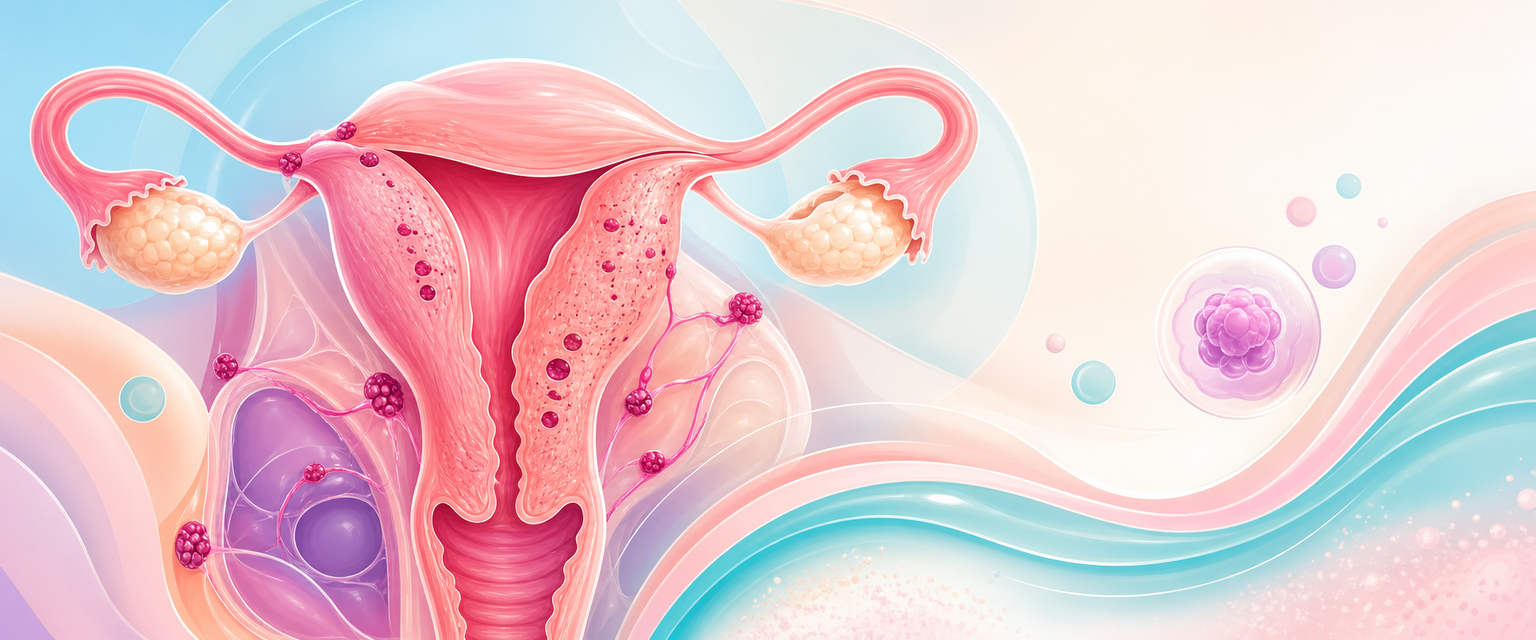
In one line
Endometriosis is a chronic, oestrogen-dependent, progesterone-resistant inflammatory disease in which endometrium-like tissue outside the uterus drives pelvic pain and subfertility; adenomyosis is its intramyometrial cousin. The consultant task is to treat the symptom the woman actually has — pain or fertility, rarely both at once with the same intervention — and to recognise that the modern diagnosis is clinical and imaging-based, that medical suppression is first-line and empirical, and that surgery for an endometrioma always costs ovarian reserve.
The disease-mechanism groundwork is covered at endometriosis pathophysiology; the focus here is the consultant decision — defending a management plan from the evidence and the South African resource reality.
Mechanism & pathophysiology
No single theory accounts for endometriosis, and a candidate who offers only retrograde menstruation has misread the disease. The mechanism is a convergence of an anatomical seeding event, a permissive host, and an autonomous lesion biology.
- Sampson's retrograde menstruation remains the dominant explanation for pelvic disease: viable endometrial fragments reflux through the tubes and implant on peritoneum, ovary and pouch of Douglas. It is anatomically persuasive — the gravity-dependent and left-sided predilection (the sigmoid shelters the left adnexa) fits — but it cannot be the whole story, because retrograde menstruation occurs in up to 90% of menstruating women while only ~10% develop endometriosis. The reflux is necessary, not sufficient.
- Coelomic metaplasia (Meyer) explains disease where reflux cannot reach — the pleura, and the rare cases in women without a functioning uterus — by proposing that peritoneal mesothelium, sharing a coelomic-epithelial origin with the Müllerian tract, transforms into endometrium-like tissue under hormonal or inflammatory stimulus.
- Stem/progenitor-cell and lymphovascular dissemination account for distant deposits (umbilical, thoracic, even cerebral), positing that bone-marrow-derived or endometrial progenitor cells circulate and engraft.
- The immune and inflammatory defect is what converts seeding into established disease. Peritoneal macrophages in affected women clear refluxed tissue poorly and instead secrete a pro-inflammatory, pro-angiogenic milieu (IL-1, IL-6, IL-8, TNF-α, VEGF) that lets implants survive, vascularise and incite the nociceptor sprouting and neuroangiogenesis underlying the pain.
Two molecular features unify the picture and dictate treatment. First, the lesions are oestrogen-dependent and locally oestrogen-generating: ectopic stroma over-expresses aromatase (absent in normal endometrium) and is deficient in 17β-hydroxysteroid dehydrogenase type 2, so it both synthesises oestradiol locally and fails to inactivate it — a self-amplifying loop, with COX-2-driven prostaglandin E2 further upregulating aromatase. This is why every effective hormonal therapy works by lowering or opposing oestrogen, and why aromatase inhibitors have a niche in refractory disease. Second, the lesions are progesterone-resistant: progesterone-receptor (chiefly PR-B) expression is blunted, so the normal progesterone brake on endometrial proliferation fails. Progesterone resistance is the reason a progestogen suppresses some women beautifully and barely touches others, and the conceptual basis for the whole disease behaving like an unopposed-oestrogen state in miniature.
The pain itself has its own pathophysiology, and understanding it explains why excising lesions does not always abolish symptoms. Early pain is nociceptive and cyclical, driven by prostaglandins and inflammatory mediators; with time the lesions recruit their own nerve supply (neuroangiogenesis), and persistent peripheral input sensitises the dorsal horn and central pain pathways. Once central sensitisation is established, pain becomes constant, spreads beyond the pelvis, and persists despite complete lesion clearance — which is why a fixated surgical answer ("find and cut all of it") fails the woman whose pain has become a chronic-pain syndrome, and why pain-medicine input, not more surgery, is sometimes the right escalation.
The disease is not one phenotype but three, with different biology and surgery:
- Superficial peritoneal endometriosis — implants on peritoneal surfaces; the commonest form, the one whose pain correlates worst with what the eye sees at laparoscopy.
- Ovarian endometrioma ("chocolate cyst") — a pseudocyst lined by endometriotic tissue and filled with altered (haemosiderin-laden) blood. Its wall is inseparable from ovarian cortex, which is why excising it removes follicles.
- Deep infiltrating endometriosis (DIE) — nodular disease penetrating >5 mm beneath the peritoneum into the uterosacral ligaments, rectovaginal septum, bowel, bladder or ureter. It behaves more like a fibrotic, adenomyosis-like lesion than a surface implant, distorts anatomy, and is the disease that obstructs ureters and infiltrates rectum — the one that demands a multidisciplinary surgical team.
Adenomyosis is endometrium-like glands and stroma within the myometrium, surrounded by reactive smooth-muscle hyperplasia. The favoured mechanism is invagination of the basal endometrium across a disrupted endometrial–myometrial junctional zone, with the same oestrogen-dependence and progesterone resistance as endometriosis, plus dysfunctional uterine peristalsis. Mechanical and inflammatory disruption of the junctional zone — by tissue-injury-and-repair at the basalis, and plausibly by prior uterine instrumentation or pregnancy — is thought to initiate the invagination, after which the ectopic foci provoke the surrounding myometrial hyperplasia that makes the uterus bulky and the junctional zone thick on imaging. The result is a globular, tender, often symmetrically enlarged uterus producing heavy menstrual bleeding and progressive dysmenorrhoea, and increasingly recognised as a contributor to subfertility and adverse obstetric outcomes through impaired junctional-zone peristalsis and implantation. The two diseases overlap heavily — a large share of women with endometriosis have coexistent adenomyosis on imaging — and that coexistence partly explains why some women fail conservative endometriosis surgery: the uterus itself is a pain generator that pelvic excision never addressed.
Assessment
Diagnosis has shifted decisively in the last decade: laparoscopy is no longer the mandatory gold standard, and a confident clinical-plus-imaging diagnosis now starts treatment without an operation. The reasoning is twofold — laparoscopy carries surgical risk and a negative laparoscopy does not exclude disease (microscopic and non-pigmented lesions are missed), and delaying empirical treatment until a diagnostic operation prolongs the already notorious diagnostic delay (typically years from symptom onset). Both ESHRE 2022 and NICE NG73 now support diagnosis on a characteristic symptom pattern and imaging, reserving surgery for treatment or for diagnostic uncertainty.
The symptom pattern is the diagnosis until proven otherwise:
- Cyclical, then chronic, pelvic pain — initially perimenstrual, progressing to constant pain as central sensitisation sets in. Pain that has lost its cyclicity signals a chronic-pain syndrome, not necessarily worse lesions.
- Secondary dysmenorrhoea that is progressive and increasingly refractory to simple analgesia.
- Deep dyspareunia — particularly with uterosacral or rectovaginal DIE.
- Cyclical bowel or bladder symptoms (dyschezia, haematochezia, dysuria, cyclical haematuria) — pointers to deep disease infiltrating rectum, bladder or ureter.
- Subfertility — by adhesions and distorted anatomy, impaired oocyte quality and a hostile inflammatory peritoneal environment.
Examination is often unremarkable in superficial disease, but the signs of deep disease are specific and worth eliciting deliberately: a fixed, retroverted uterus, tender nodularity or thickening of the uterosacral ligaments and rectovaginal septum on bimanual and rectovaginal examination, an adnexal mass (endometrioma), and visible blue/red lesions in the posterior fornix. A bulky, tender, globular uterus suggests adenomyosis.