In one line
Uterine fibroids are benign monoclonal smooth-muscle tumours whose symptoms — heavy menstrual bleeding, bulk effects and reproductive consequences — are predicted far better by the FIGO 0–8 location of the fibroid than by its size, so the consultant task is to match the intervention to the symptom and to the woman's fertility intent, sparing the uterus wherever that is what she values and reserving hysterectomy for the woman who has completed her family and wants the problem gone for good.
The bleeding mechanism and first-line medical control are developed in depth at Heavy menstrual bleeding; the groundwork on pathology, basic classification and presentation is assumed from the Intermediate chapter fibroids. The focus here is the consultant decision — choosing between, and defending, the uterine-sparing and definitive options, and managing the tumour through pregnancy and the perimenopause.
Mechanism & pathophysiology
Each fibroid is an independent monoclonal proliferation: a single myometrial smooth-muscle cell acquires a driver lesion and clonally expands, which is why a woman commonly carries multiple genetically distinct tumours rather than one disease that has spread. The commonest driver, present in roughly 50–85% of leiomyomas, is a mutation in MED12 (a subunit of the Mediator transcription-regulatory complex); the next, in most MED12-wild-type tumours, is overexpression of the architectural transcription factor HMGA2, usually through a 12q14–15 rearrangement. These are largely mutually exclusive events. A clinically important minority are fumarate hydratase (FH)-deficient, the uterine manifestation of hereditary leiomyomatosis and renal cell cancer (HLRCC) — a woman with multiple early-onset, often symptomatic fibroids and cutaneous leiomyomas or a family history of aggressive renal cancer warrants genetic referral, because the renal tumours are lethal and screenable.
Whatever the driver, the tumour is steroid-hormone-dependent. Fibroid tissue overexpresses both oestrogen and progesterone receptors relative to normal myometrium, and — counter to the old teaching that they are purely oestrogen-driven — progesterone is the dominant proliferative signal: progesterone receptor activation drives mitosis and survival, oestrogen largely by upregulating that progesterone-receptor machinery. This is the rationale for every effective medical therapy. Removing the steroid drive (GnRH analogues), blocking the progesterone receptor (selective progesterone-receptor modulators), or suppressing the gonadal axis with controlled add-back (GnRH antagonist combinations) all shrink fibroids and stop bleeding; the corollary is that fibroids characteristically grow through the reproductive years, can enlarge in pregnancy, and regress after the menopause as the hormonal substrate falls away — which itself is part of the management calculus in the woman near 50.
The bulk of a fibroid is not cells but extracellular matrix — a disordered, collagen-rich, stiff matrix that the tumour cells actively deposit and remodel. This matters mechanically: the stiffness and the slow matrix turnover explain why fibroids are firm, why they outgrow their blood supply and undergo degeneration (hyaline, cystic, calcific, and the painful red/carneous degeneration of pregnancy, in which a rapidly enlarging fibroid infarcts), and why purely cytoreductive drugs shrink them only so far.
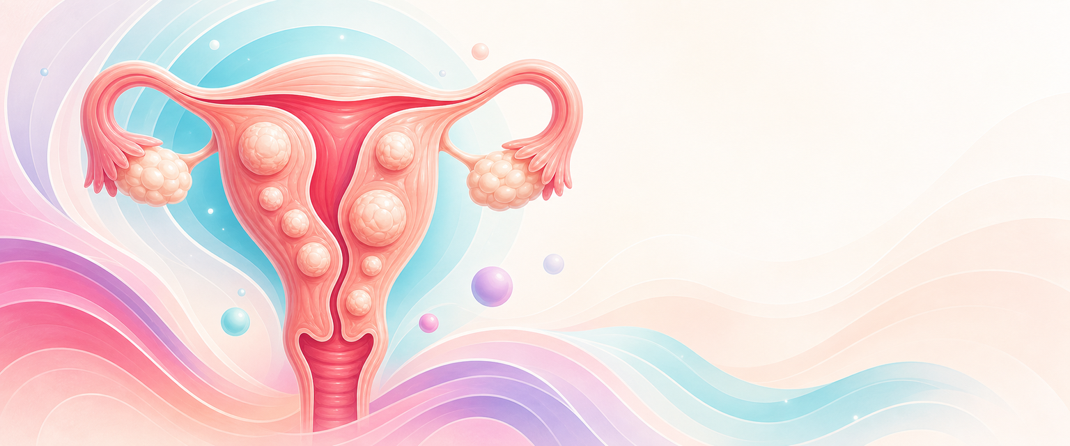
The clinical syndromes follow directly from where this matrix sits, codified in the FIGO leiomyoma subclassification (types 0–8) nested within PALM-COEIN:
- Submucosal (types 0–2) distort the endometrial cavity. Type 0 is wholly intracavitary on a stalk; type 1 has <50% intramural extension; type 2 has ≥50%. These are the fibroids that cause heavy menstrual bleeding out of proportion to their size — by increasing and distorting the endometrial surface, dysregulating local vasoactive and clotting factors, and impairing myometrial contraction — and the ones that impair implantation and raise miscarriage risk. A small submucosal fibroid can dominate the clinical picture; a large subserosal one may be silent.
- Intramural (types 3–4) sit within the myometrium. Type 3 contacts the endometrium without distorting it; type 4 lies entirely within the myometrium, touching neither surface. They contribute to bleeding and bulk, and their effect on fertility is the contested middle ground (below).
- Subserosal (types 5–7) reach or project beyond the serosa. Type 5 is at least half intramural but contacts the serosa; type 6 is less than half intramural; type 7 is pedunculated. These produce bulk and pressure symptoms, such as urinary frequency, retention, hydronephrosis from ureteric compression, constipation, abdominal distension and the dragging discomfort of a heavy pelvic mass, rather than bleeding.
- Type 8 is "other" — cervical, or parasitic fibroids that have acquired an extra-uterine blood supply.
- Hybrid lesions touching both endometrium and serosa are written as two numbers (e.g. 2–5).
The fertility and obstetric mechanisms are the same anatomy applied to pregnancy, and they sort cleanly by FIGO type. Submucosal (cavity-distorting) fibroids reduce implantation and live-birth rates and increase miscarriage, and their resection improves outcomes — the one situation where the fertility benefit of treatment is reasonably secure. Subserosal fibroids do not measurably affect fertility. The genuinely contested ground is the intramural fibroid that does not distort the cavity: the meta-analytic signal is a modest reduction in implantation and live birth, but the evidence that removing such a fibroid restores fertility is weak, so myomectomy here is a judgement call, not a reflex. In an established pregnancy, fibroids raise the risks of malpresentation (a lower-segment fibroid displaces the presenting part; OR ~2.7), obstructed labour, placental abruption (OR ~2.6) and praevia (OR ~2.2), preterm birth (OR ~1.5), caesarean delivery (OR ~2.6), and postpartum haemorrhage (OR ~3) from an impaired contractile uterus. Red degeneration — acute, severe, localised pain with low-grade fever and a tender enlarging fibroid in the second trimester, occurring in roughly 1 in 12 pregnant women with fibroids — is the characteristic pregnancy event and is managed conservatively.
The diagnosis that haunts this whole field is the rare uterine leiomyosarcoma. It is not a fibroid that has "turned malignant" — the two are largely distinct from the outset, and malignant transformation of a benign fibroid is vanishingly rare. The problem is that no current imaging or clinical feature reliably distinguishes a benign fibroid from a sarcoma before histology, and a leiomyosarcoma can masquerade as a "growing fibroid". This diagnostic uncertainty is the engine of the morcellation controversy addressed below: an operation that fragments the specimen will, in the unlucky woman with an occult sarcoma, disseminate it.
Assessment
- History. Characterise the dominant symptom, because it predicts both the FIGO type and the right intervention. Bleeding — heavy, prolonged, with flooding/clots, fatigue and the symptoms of iron deficiency — points to a submucosal or large intramural lesion. Bulk/pressure — abdominal swelling, urinary frequency or incomplete emptying, constipation, dyspareunia, a palpable mass — points to subserosal or large intramural disease. Ask specifically about subfertility and adverse obstetric history, and about the woman's reproductive intent, because that single fact reorganises the whole management plan. Acute severe pain suggests degeneration or torsion of a pedunculated fibroid.
- Examination. Abdominal palpation may reveal a firm, irregular, non-tender mass arising from the pelvis and (unlike an ovarian mass) usually one you cannot get below; bimanual examination finds an enlarged, mobile, irregular uterus that moves with the cervix — the bedside discriminator from an adnexal mass, which moves independently. Quantify the size in weeks-of-gestation equivalent and look for the pallor and tachycardia of anaemia.
- Transvaginal ultrasound is the first-line investigation: it confirms fibroids, sizes and counts them, and locates them well enough to assign FIGO type in most cases. Saline-infusion sonography (sonohysterography) is the key refinement for submucosal lesions — distending the cavity defines the type 0/1/2 boundary precisely, which is exactly the information that decides whether a hysteroscopic resection is feasible.
- Assigning the FIGO type — the language that drives the plan. Imaging exists to place each fibroid in the FIGO leiomyoma subclassification, because the type, not the size, selects the procedure: