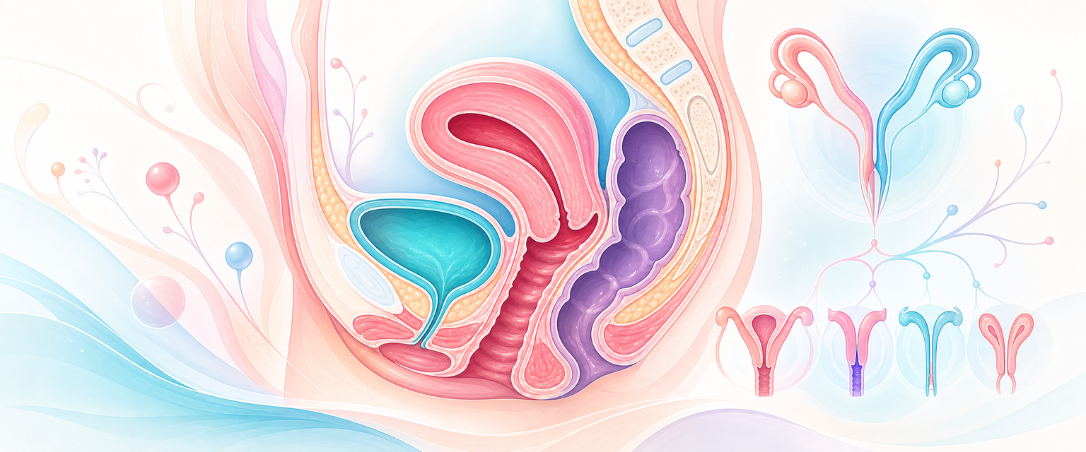
In one line
Müllerian anomalies are a developmental spectrum from a fundal septum to complete uterovaginal agenesis; the consultant tasks are to separate an obstructive anomaly (an adolescent emergency of trapped menstrual blood) from a reproductive one (recurrent loss or preterm birth), to image the kidneys in every case because the urinary tract shares the embryology, and to resist the single most over-performed operation in the field — hysteroscopic resection of a uterine septum, which the only randomised trial failed to show improves live birth.
Mechanism & pathophysiology
The female genital tract is built by three processes that fail independently, and which anomaly a woman has is read off which process failed.
The paired paramesonephric (Müllerian) ducts form alongside the mesonephric (Wolffian) ducts on the urogenital ridge. In the genetic female, the absence of anti-Müllerian hormone lets the Müllerian ducts persist while the Wolffian ducts regress. Each duct elongates caudally, the two fuse in the midline to form the uterus, cervix and upper vagina, and the intervening septum is then resorbed to leave a single cavity. The lower vagina has a separate origin: it canalises from the urogenital sinus, and the sinovaginal bulbs meet the descending Müllerian tissue at the vaginal plate, which then hollows out. Three sequential steps — formation, fusion, resorption — and a failure at each produces a recognisable lesion:
- Agenesis/hypoplasia (formation fails). One duct fails to form (a true unicornuate uterus, sometimes with a rudimentary contralateral horn) or both fail (Müllerian aplasia — the uterovaginal component of MRKH, Mayer–Rokitansky–Küster–Hauser syndrome). The ovaries, derived from the genital ridge rather than the ducts, are unaffected — which is why an MRKH patient has normal pubertal development and normal endocrine function with an absent uterus and a blind or absent vagina.
- Fusion failure. The two horns never come together: a complete failure gives uterus didelphys (two cervices, often a longitudinal vaginal septum); a partial one gives a bicornuate uterus (two horns sharing a single cervix). The defining feature is an externally cleft fundus — fusion was the step that failed.
- Resorption failure. Fusion succeeded but the midline septum was never removed, leaving a septate uterus with a smooth, intact external fundal contour and a divided cavity. This is the commonest anomaly and the one with the worst miscarriage record, and the septum itself — relatively avascular, fibromuscular, a poor implantation surface — is what drives the reproductive harm.
The single most important corollary follows from the duct's neighbour. The mesonephric (Wolffian) duct directs ureteric budding and metanephric induction, so a Müllerian malformation is frequently accompanied by a urinary-tract one — most classically unilateral renal agenesis on the same side as the obstructed or absent Müllerian structure. In a Chinese tertiary series of 444 reproductive-tract anomalies, extragenital malformations accompanied 43.5%, the urinary tract was involved in 30.6%, and renal agenesis on the obstructed side was present in 100% of oblique-vaginal-septum (OHVIRA) cases (Su 2024). Imaging the kidneys is therefore not optional thoroughness; it is part of making the diagnosis.
Two mechanistic points separate the consultant from the trainee. First, obstruction is a function of anatomy plus a functioning endometrium. A non-communicating cavity (a rudimentary horn with endometrium, an obstructed hemivagina, an imperforate hymen) is silent until menarche, then fills cyclically — haematometra, haematocolpos, retrograde menstruation and its sequel, endometriosis. The clock starts at menarche, which is why these present in adolescence. Second, a longitudinal vaginal septum or didelphys does not by itself cause subfertility; the harm in unification defects is obstetric — preterm birth and malpresentation from a smaller, less distensible cavity — not a failure to conceive.
The septum's reproductive toxicity is worth understanding at tissue level, because it is the rationale that the randomised data later undercut. The fundal septum is relatively avascular fibromuscular tissue with a thin, poorly vascularised endometrial covering and altered local progesterone receptivity — an implantation surface that supports a pregnancy badly. An embryo implanting on the septum sits on a poor blood supply; the proposed mechanisms for the excess miscarriage are reduced decidual perfusion, distorted cavity geometry and impaired distension. That is a coherent story, and it is exactly why "find a septum, cut a septum" felt self-evidently correct for decades — the lesson of this topic is that a plausible mechanism is not a substitute for a trial.
MRKH itself sits at the formation end as a more specific entity. It is Müllerian aplasia in a 46,XX woman with normal ovarian function, occurring in roughly 1 in 4,500–5,000 female births. Type 1 (isolated) is uterovaginal aplasia alone; type 2 (MURCS association) adds renal, skeletal (especially vertebral/Klippel–Feil) and occasionally cardiac and hearing anomalies — which is why the MRKH workup is never just a pelvic scan but a renal, spinal and cardiac survey. The genetics are heterogeneous and mostly sporadic; no single causative gene explains the majority, so counselling is about phenotype and associated anomalies rather than a clean Mendelian recurrence risk.
Assessment
The presentation tells you which arm of the spectrum you are in before any imaging.
- The obstructed adolescent. Primary amenorrhoea (or cryptomenorrhoea — cyclical pain with no visible bleeding) with worsening cyclical lower abdominal pain, a perimenstrual mass, sometimes acute urinary retention. An imperforate hymen gives a bulging bluish membrane at the introitus; a transverse vaginal septum or distal atresia gives cyclical pain with a normal-looking introitus and a short or blind vagina — a critical distinction, because incising what looks like a hymen but is a high transverse septum is dangerous. OHVIRA (obstructed hemivagina + ipsilateral renal agenesis, classically with didelphys) gives cyclical pain despite normal menstruation, because one of two systems is obstructed — a paratubal/paravaginal mass with regular periods is OHVIRA until proven otherwise.
- The woman with normal periods and obstetric trouble. Recurrent first-trimester miscarriage, mid-trimester loss, preterm birth or a persistent transverse/oblique lie point to a septate or unification defect found on investigation rather than examination.
- Primary amenorrhoea with normal secondary sexual characteristics. MRKH: normal breast and pubic-hair development (ovaries intact), a blind vaginal dimple, absent or rudimentary uterus on ultrasound. The differential that must be excluded is complete androgen insensitivity syndrome — also amenorrhoea with a blind vagina, but a 46,XY karyotype, absent/scant pubic hair and intra-abdominal testes. A karyotype is mandatory before MRKH is settled; the management, gonadal-cancer risk and counselling diverge completely. This overlaps the disorders-of-sex-development workup covered at Disorders of sex development and the pubertal differential at Puberty and its disorders.
- The incidental finding. A septate or arcuate uterus found at caesarean, laparoscopy or routine scan in an asymptomatic, normally fertile woman — a finding that frequently needs no intervention, and over-treating it is a recognised harm.
Investigation is led by imaging, and the reference standards are specific:
- 3D transvaginal ultrasound is the practical first-line and, in expert hands, approaches MRI for the septate-versus-bicornuate distinction because the coronal plane shows the fundal contour. It is operator-dependent, which matters in a service where 3D probes and the skill to use them are concentrated in a few centres.
- MRI is the reference standard for the full anatomy: it shows the external fundal contour (the feature that separates septate from bicornuate), the presence and content of a rudimentary horn, the level of a vaginal septum, cervical and vaginal anatomy, and associated renal position. The septate-versus-bicornuate call is the one that changes management most and is the one a 2D scan or hysterosalpingogram (which sees only the cavity, never the external contour) cannot make — an HSG showing two cavities is equally compatible with a septate and a bicornuate uterus.
- Renal tract imaging in every confirmed anomaly — ultrasound at minimum, because of the shared embryology and the 100% renal-agenesis association in OHVIRA.
- Karyotype where MRKH is suspected (to exclude CAIS) and where the phenotype is ambiguous.