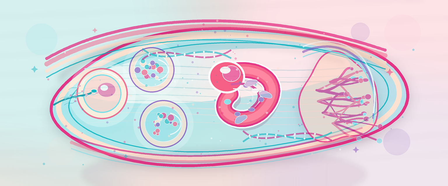
Early and Systematic Embryological Development
Early embryology is timed patterning. The body plan is not assembled randomly; it follows a sequence of fertilisation, cleavage, implantation, bilaminar disc formation, gastrulation, folding, neurulation, organogenesis and fetal growth. A candidate who knows the timeline can reason through teratogens, congenital anomalies, miscarriage, twin chorionicity, DSD and abnormal placentation.
The high-yield timeline is:
- day 0: fertilisation in the ampulla of the tube;
- days 1 to 3: cleavage divisions;
- day 3 to 4: morula;
- day 5: blastocyst;
- day 6 to 7: implantation begins;
- week 2: bilaminar disc, amnion, yolk sac and early trophoblast lacunae;
- week 3: gastrulation and trilaminar disc;
- weeks 3 to 8: organogenesis;
- week 9 onward: fetal growth and functional maturation.
The exact dates vary slightly between descriptions, but the sequence is what matters.
| Time | Developmental event | Clinical meaning |
|---|---|---|
| Day 0 | Fertilisation | Restores diploidy; errors can cause triploidy or failed development |
| Days 1-3 | Cleavage | Rapid mitosis; early loss is often chromosomal |
| Day 5 | Blastocyst | Trophoblast and inner cell mass separate |
| Days 6-7 | Implantation | Ectopic pregnancy and abnormal implantation become possible |
| Week 2 | Bilaminar disc | Amnion, yolk sac and trophoblast lacunae develop |
| Week 3 | Gastrulation | Three germ layers form; severe body-plan errors occur here |
| Weeks 3-8 | Organogenesis | Highest risk window for structural teratogenesis |
| Week 9 onward | Fetal period | Growth and functional maturation dominate |
The key is to link time to vulnerability. A harmful exposure before implantation may cause loss without a structural malformation. An exposure during organogenesis can alter structure. An exposure later may alter growth, function, endocrine programming or neurodevelopment.
Developmental Time Versus Gestational Age
Embryology often counts from fertilisation, while obstetrics usually counts from the last menstrual period. The obstetric gestational age is roughly two weeks more than fertilisation age in a regular 28-day cycle. This matters when counselling about exposure timing.
| Embryology age | Approximate obstetric age | Developmental meaning |
|---|---|---|
| Fertilisation day 0 | 2 weeks | Zygote begins |
| Week 3 after fertilisation | 5 weeks | Gastrulation, neurulation, early heart development |
| Weeks 3-8 after fertilisation | 5-10 weeks | Organogenesis |
| Week 9 onward after fertilisation | 11 weeks onward | Fetal growth and maturation |
When a patient says she took a drug at "six weeks pregnant", an O&G answer must clarify whether that is six weeks by last menstrual period. That usually places the embryo around four weeks after fertilisation, a period of active early organogenesis.
Fertilisation
Fertilisation usually occurs in the ampulla of the uterine tube. It is a process, not a single collision. Sperm must undergo capacitation in the female reproductive tract, which makes them capable of the acrosome reaction. The acrosome reaction helps sperm penetrate the corona radiata and zona pellucida. Fusion with the oocyte membrane triggers oocyte activation and cortical granule release, reducing the chance of polyspermy.
The oocyte completes meiosis II after sperm entry. Male and female pronuclei form, DNA is prepared, and the first mitotic division follows. This restores diploidy and starts embryonic development.
Fertilisation failures explain important clinical outcomes:
- failure to fertilise gives infertility;
- polyspermy can cause triploidy;
- abnormal parental genomic contribution can cause molar pregnancy;
- tubal transport failure can produce ectopic pregnancy;
- early chromosomal errors can produce miscarriage.
It is worth understanding the mechanism behind two of those outcomes, because they are common Primary stems. Triploidy (three full chromosome sets, 69 chromosomes) most often arises when two sperm fertilise one egg, or one diploid sperm fertilises a normal egg; the extra set is then paternal (this pattern is associated with a partial hydatidiform mole and a triploid fetus). Less commonly the extra set is maternal, from a failure of meiosis that leaves the egg diploid. The balance of parental genomes matters because of genomic imprinting: certain genes are expressed only from the maternally or only from the paternally inherited copy. An excess of paternal genome drives trophoblast overgrowth, which is why molar pregnancies — where the genetic contribution is paternal-skewed — produce the abnormal, hydropic trophoblast and high hCG that define gestational trophoblastic disease. Primary only needs the principle: it is not just how many chromosome sets, but whose, that shapes the outcome.
The sperm contributes more than DNA, and the oocyte contributes more than chromosomes. The oocyte provides cytoplasm, mitochondria, stored RNAs and proteins needed for early cleavage. This is why oocyte quality is so important in early embryo competence. The early embryo initially runs on maternal stores before embryonic genome activation takes over — the point at which the embryo's own genome, rather than inherited maternal transcripts, begins to direct development.
Cleavage, Morula and Blastocyst
Cleavage divisions are rapid mitotic divisions inside the zona pellucida. The total conceptus does not grow much at first; cells become smaller blastomeres. This allows the embryo to travel through the tube without implanting too early.
The morula is a compact ball of cells. As fluid enters, a blastocyst forms. The blastocyst has two major components:
- trophoblast, which contributes to placenta and membranes;
- inner cell mass, which contributes to the embryo proper and some extraembryonic structures.
The blastocyst must hatch from the zona pellucida before implantation. If hatching or tubal transport is abnormal, implantation may fail or occur outside the uterine cavity.
Early lineage decisions begin here:
| Blastocyst component | Main derivative | O&G importance |
|---|---|---|
| Trophoblast | Placenta and chorionic structures | Implantation, hCG, GTD, placental disease |
| Embryoblast / inner cell mass | Embryo proper and some extraembryonic tissues | Body plan and organogenesis |
| Blastocyst cavity | Fluid-filled space organising implantation stage | Hatching and implantation readiness |
This is the first major separation between fetal support tissue and embryo proper. It also explains why a placental genetic result can differ from the fetus if later mosaicism or lineage restriction occurs.
Implantation and Early Placental Development
Implantation requires a receptive endometrium and competent trophoblast. The trophoblast differentiates into two layers with different jobs. The cytotrophoblast is an inner layer of dividing single cells; it is the proliferative reservoir. The syncytiotrophoblast is an outer multinucleated layer formed by fusion of cytotrophoblast cells, with no internal cell boundaries. The syncytiotrophoblast is the invasive, hormone-producing and transport surface that sits directly against maternal blood. Keeping these two layers straight is worth the effort, because almost every placental disease is a disorder of one of them.
As the syncytiotrophoblast invades the endometrium it erodes maternal capillaries, and fluid-filled spaces called lacunae open within it. These lacunae fill with maternal blood and become confluent — they are the precursor of the intervillous space, the lake of maternal blood that the placenta will eventually bathe its villi in. This is the structural beginning of the maternal–fetal exchange interface.