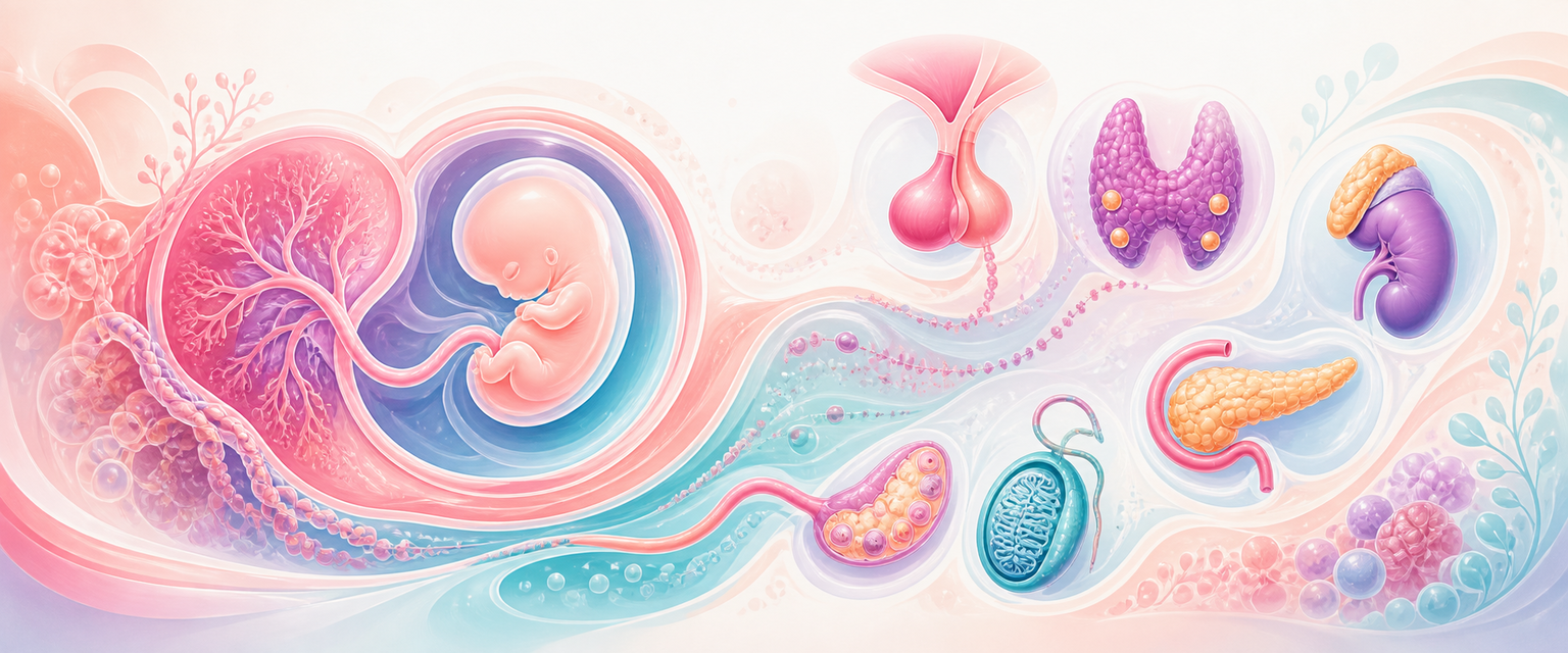
Endocrine Organ Development
Start with one idea: an endocrine organ is a piece of tissue that makes a chemical message and posts it into the blood. Every gland in this chapter is built from the same three early embryonic sheets — ectoderm, mesoderm and endoderm — plus the migratory neural crest and, uniquely in pregnancy, the trophoblast. None of these tissues "knows" it will become an endocrine organ; each is simply at a particular place at a particular time, receives the right signals, and differentiates. Once you hold that, the whole chapter becomes a single question asked organ by organ: which sheet, which place, which signal, and what message does the finished gland post?
Endocrine embryology matters in O&G because pregnancy is an endocrine stress test. Thyroid disease affects fetal brain development. Diabetes affects organogenesis and fetal growth. Congenital adrenal hyperplasia causes virilised 46,XX external genitalia. Pituitary disease affects puberty, ovulation, lactation and adrenal reserve. The placenta is itself a temporary endocrine organ, and the fetal adrenal gland supplies steroid precursors that the placenta cannot make alone.
The endocrine system is not one embryological organ. It is a network built from different germ layers and then connected by hormones. Three principles run through everything below, so meet them now and the tables will read as worked examples rather than lists:
- Origin predicts anatomy. Where a gland forms — and the path it migrates — predicts the structural anomalies you will see (an ectopic, a cyst, a duct remnant).
- Hormone output predicts presentation. What the finished gland secretes predicts the clinical picture when it fails or over-functions.
- Timing predicts the lesion. The same insult (hyperglycaemia, hypothyroxinaemia, an androgen excess) does completely different things depending on whether it strikes during organ formation or during later growth.
| Organ | Main embryological origin | O&G relevance |
|---|---|---|
| Anterior pituitary | Oral ectoderm from Rathke pouch | Puberty, ovulation, prolactin, adrenal and thyroid axes |
| Posterior pituitary | Neuroectoderm from hypothalamus | Oxytocin, vasopressin, gestational diabetes insipidus logic |
| Pineal gland | Neuroectoderm (roof of third ventricle) | Melatonin; circadian and reproductive-axis modulation |
| Thyroid follicular cells | Endoderm from floor of primitive pharynx | Fetal neurodevelopment, maternal thyroid disease |
| Parafollicular C cells | Neural crest/ultimobranchial body contribution | Calcitonin, medullary thyroid cancer context |
| Parathyroids | Third and fourth pharyngeal pouches | Calcium physiology in pregnancy/lactation |
| Adrenal cortex | Mesoderm near urogenital ridge | Cortisol, aldosterone, fetal DHEAS, CAH |
| Adrenal medulla | Neural crest | Catecholamines, stress response, phaeochromocytoma bridge |
| Pancreatic islets | Endoderm from pancreatic buds | Diabetes, fetal hyperinsulinaemia |
| Gonads | Urogenital ridge + germ cells | Steroids, gametes, DSD and PMOS |
| Placenta | Trophoblast and extraembryonic tissues | hCG, progesterone, hPL, placental CRH, fetal-placental steroidogenesis |
Read the middle column as germ-layer bookkeeping. Two endocrine glands come from ectoderm (the anterior pituitary from oral ectoderm, the posterior pituitary and pineal from neuroectoderm). Three come from endoderm (thyroid follicular cells, parathyroids and pancreatic islets, all derivatives of the primitive gut tube and its pharyngeal pouches). The adrenal cortex and gonadal steroid cells come from mesoderm of the urogenital ridge. The adrenal medulla and thyroid C cells come from neural crest. The placenta is the outlier — trophoblast, an extraembryonic tissue that exists only in pregnancy. The same insult therefore rarely hits "the endocrine system" as a block; it hits whichever germ layer is differentiating at that moment.
The exam principle is that origin predicts anatomy, while hormone output predicts clinical presentation.
The Endocrine Embryology Map
Endocrine organs develop in different places, but they become clinically connected through axes. A pituitary defect may present as delayed puberty. A thyroid-development defect may present as neonatal hypothyroidism. A pancreatic beta-cell environment shaped by maternal glucose may present as macrosomia. A fetal adrenal enzyme block may present as ambiguous genitalia. The candidate must therefore hold two maps at once:
| Map | What it explains | Example |
|---|---|---|
| Embryological origin | Where the organ forms and what anomalies follow | Thyroglossal duct cyst follows thyroid descent |
| Hormone axis | What physiological failure looks like | Low cortisol drives high ACTH and adrenal hyperplasia |
| Timing | When fetal development is vulnerable | Hyperglycaemia during organogenesis causes malformations; later hyperglycaemia causes overgrowth |
| Tissue response | Which tissues respond to the hormone | Androgen-sensitive external genitalia virilise in 46,XX CAH |
Approximate Timing Anchors
| Developmental window | Endocrine event | O&G meaning |
|---|---|---|
| Early first trimester | Pituitary primordia, thyroid descent, pancreatic buds, gonadal ridges | Teratogenic and metabolic injury can affect organogenesis |
| Late first trimester | Fetal thyroid begins concentrating iodine and producing hormone; gonadal differentiation becomes clearer | Maternal thyroid hormone still matters; DSD patterns begin to declare themselves |
| Second trimester | Fetal adrenal fetal zone and placental steroid unit become prominent | Oestriol biology depends on fetal adrenal, fetal liver and placenta |
| Third trimester | Fetal adrenal cortisol rise, lung/gut/liver maturation, pancreatic insulin growth effects | Steroids, diabetes control and fetal growth patterns become clinically visible |
| Neonatal period | Placental hormone withdrawal, fetal adrenal-zone involution, glucose transition | Neonatal hypoglycaemia, salt-wasting CAH and congenital hypothyroidism must be detected |
Do not memorise exact days at the expense of the concept: early pregnancy is organ formation; late pregnancy is growth, maturation and endocrine adaptation.
Pituitary Development
The pituitary has dual origin. The anterior pituitary develops from oral ectoderm that grows upward as Rathke pouch. The posterior pituitary develops from a downward extension of neuroectoderm from the diencephalon/hypothalamus. These two tissues meet and form one gland with two very different functions.
| Pituitary part | Origin | Hormones/function |
|---|---|---|
| Anterior pituitary | Rathke pouch, oral ectoderm | ACTH, TSH, LH, FSH, prolactin, GH |
| Posterior pituitary | Neurohypophyseal downgrowth from hypothalamus | Stores/releases oxytocin and vasopressin made in hypothalamus |
| Pituitary stalk | Neuroectodermal connection | Portal blood and neural continuity |
This dual origin explains several clinical patterns:
- pituitary developmental lesions may affect anterior hormone production, posterior hormone release or both;
- stalk disruption can cause diabetes insipidus or altered prolactin control;
- hypothalamic disease can suppress GnRH pulses without primary pituitary failure;
- lactation depends on prolactin secretion and oxytocin-mediated milk ejection.
At Primary level, do not learn the pituitary as a floating "master gland". Learn it as a developmental meeting point between brain and endocrine tissue.
Pituitary Pattern Recognition
| Developmental problem | Likely endocrine pattern | O&G bridge |
|---|---|---|
| Rathke pouch/anterior pituitary defect | Low ACTH, TSH, LH/FSH, GH or prolactin depending extent | Delayed puberty, amenorrhoea, infertility, adrenal crisis risk |
| Stalk interruption | Diabetes insipidus, hyperprolactinaemia or combined pituitary dysfunction | Polyuria, lactation failure or menstrual dysfunction |
| Hypothalamic GnRH pulse disruption | Low gonadotrophins with structurally normal pituitary | Functional hypothalamic amenorrhoea, delayed puberty, chronic illness |
| Pituitary mass effect later in life | Hyperprolactinaemia or hypopituitarism | Anovulation, galactorrhoea, visual symptoms |
For exams, separate production from release. Oxytocin and vasopressin are made in hypothalamic neurons and released from the posterior pituitary. Prolactin is made in the anterior pituitary and is normally held back by dopamine. That difference explains why stalk lesions can raise prolactin but impair posterior-pituitary function.
Thyroid Development
The thyroid is one of the earliest endocrine glands to form. Follicular thyroid tissue begins as an endodermal thickening in the floor of the primitive pharynx near the future foramen caecum. It descends in the midline through the neck as the thyroid diverticulum, temporarily connected to the tongue by the thyroglossal duct. The duct normally disappears.