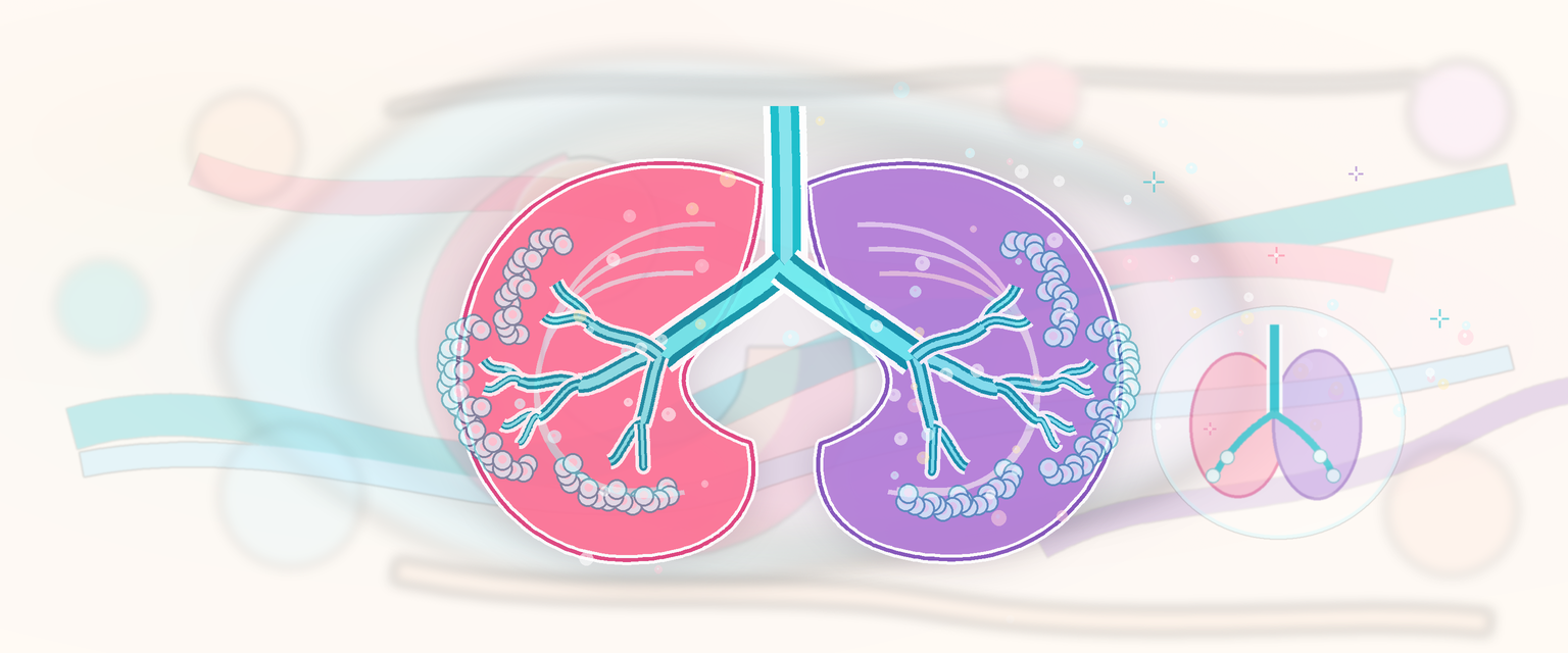
Respiratory System and Fetal Lung Development
Start from one idea and the whole chapter follows: before birth the fetal lung is a growing fluid organ that does no breathing, and the placenta does the gas exchange instead. The lung is built in liquid, supplied by the placenta, and only switches to being a gas-exchange organ in the minutes around delivery. Hold that single sentence and many O&G problems become predictable rather than memorised: preterm respiratory distress, antenatal corticosteroids, pulmonary hypoplasia after prolonged oligohydramnios, congenital diaphragmatic hernia, tracheo-oesophageal fistula, neonatal transition and persistent pulmonary hypertension all turn on either how well the lung was built in liquid or how cleanly it switched to air.
The fetal lung must achieve four things before it can work after birth:
- create a branching airway tree;
- bring capillaries close to future air spaces;
- produce surfactant;
- switch from fluid secretion to fluid absorption and air breathing at birth.
The developmental chain is:
foregut endoderm -> laryngotracheal diverticulum -> trachea and lung buds -> branching morphogenesis -> canalisation and vascularisation -> terminal sacs -> surfactant -> alveolarisation and postnatal growth
The exam skill is to convert gestational age into risk:
| Developmental window | What is being built | What the candidate should infer |
|---|---|---|
| 4-7 weeks | laryngotracheal groove, lung buds, tracheo-oesophageal separation, early lobar pattern | major airway, foregut and diaphragm-related anomalies arise early |
| 5-16/17 weeks | conducting airway branching and mesenchymal framework | no gas-exchange units; early severe oligohydramnios or small thorax can cause severe hypoplasia |
| 16-25/28 weeks | respiratory bronchioles, capillary approach, type I and type II pneumocyte differentiation | viability begins as structure and gas-exchange surface appear, but reserve remains limited |
| 24 weeks to term | saccules, surfactant accumulation, thinning of interstitium, vascular preparation | antenatal corticosteroids and neonatal surfactant can modify outcome |
| Late fetal to childhood | alveolar septation, microvascular maturation and growth | preterm birth interrupts a programme that normally continues after birth |
This is why a 24-week fetus with prolonged anhydramnios is not simply "preterm". The lung may be structurally undergrown as well as biochemically immature.
Origin of the Respiratory Tract
The lower respiratory tract develops from endoderm of the ventral foregut, surrounded by splanchnic mesoderm. The endoderm forms the epithelial lining of the larynx, trachea, bronchi and alveoli. The surrounding mesoderm forms cartilage, smooth muscle, connective tissue, pleura and pulmonary vessels.
The laryngotracheal diverticulum grows from the foregut. The tracheo-oesophageal septum separates the ventral respiratory tube from the dorsal oesophagus. If this separation is abnormal, tracheo-oesophageal fistula and oesophageal atresia patterns result.
| Tissue | Main derivative |
|---|---|
| Foregut endoderm | Respiratory epithelium, glandular lining, alveolar epithelium |
| Splanchnic mesoderm | Cartilage, smooth muscle, connective tissue, visceral pleura, vessels |
| Somatic mesoderm | Parietal pleura and body wall contribution |
This dual origin explains why airway epithelium and airway support can be affected differently.
A few anatomical anchors are worth fixing early because the rest of development is just repetition of the same branching rule. The single laryngotracheal tube divides into two primary lung buds; the right bud subdivides into three secondary buds and the left into two, which is why the mature right lung has three lobes and the left has two. The same airway sleeve elongates downward, so the trachea grows caudally and its bifurcation sits at roughly the level of the fourth thoracic vertebra by term. Branching then continues distally long after the lobar pattern is set, but genuine gas-exchange air spaces (alveoli) do not begin to appear until around the sixth month of intra-uterine life. This timing is the structural reason a very early preterm lung has airways but almost no surface to exchange gas across.
Foregut Separation Patterns
Tracheo-oesophageal anomalies are best understood as a partitioning problem, not as two unrelated tubes.
| Pattern | Developmental idea | Antenatal clue | Neonatal clue |
|---|---|---|---|
| Oesophageal atresia with distal fistula | dorsal oesophagus interrupted, distal airway-gut communication persists | polyhydramnios may occur; stomach may still be visible because air/fluid can reach stomach through fistula | choking, coughing, cyanosis with feeds; inability to pass tube; gas in abdomen |
| Pure oesophageal atresia | oesophagus interrupted without airway-gut communication | small or absent stomach more likely, polyhydramnios more likely | no distal bowel gas; feeding/aspiration risk |
| H-type fistula | narrow airway-gut communication without atresia | often missed antenatally | recurrent coughing, aspiration and chest infection with feeds |
| Laryngeal/tracheal obstruction sequence | airway lumen or outlet obstructed | enlarged echogenic lungs, flattened/inverted diaphragm, hydrops in severe cases | airway emergency if not recognised |
The scan is therefore indirect. Normal liquor and a visible stomach do not fully exclude tracheo-oesophageal fistula, especially when a distal fistula is present.
Branching Morphogenesis
The lung buds branch repeatedly to form the bronchial tree. Branching is directed by epithelial-mesenchymal signalling. The right lung forms three main lobes and the left lung two, reflecting bronchial branching and thoracic layout.
Branching is not random. It requires growth factors, extracellular matrix, mechanical stretch, fluid pressure and surrounding thoracic space. A lung trapped in a small thorax or compressed by abdominal contents will not branch and expand normally.
| Requirement for normal growth | Failure pattern |
|---|---|
| Adequate thoracic space | Pulmonary hypoplasia in congenital diaphragmatic hernia or skeletal dysplasia |
| Adequate amniotic fluid | Pulmonary hypoplasia after severe early oligohydramnios |
| Patent airways and lung liquid | Hypoplasia if airway obstruction or fluid dynamics fail |
| Fetal breathing movements | Reduced growth with neuromuscular impairment |
| Placental oxygen/substrate supply | Growth and maturation impairment |
Branching morphogenesis has a proximal-distal logic. Proximal airways need cartilage, smooth muscle and ciliated epithelium; distal air spaces need thin epithelium, surfactant-producing cells and a dense capillary network. The fetal lung is therefore patterned both as an airway tree and as a future gas-exchange surface.
| Signal/process | Simple exam meaning | If disturbed |
|---|---|---|
| FGF-type epithelial outgrowth signals | drive bud outgrowth and branching | reduced or abnormal branching |
| SHH/BMP/TGF-beta balance | restrains and shapes branching | abnormal airway pattern or over/under-branching |
| WNT and NKX2.1 lung fate signals | specify respiratory epithelial identity | severe epithelial differentiation problems |
| VEGF and vascular coupling | brings capillaries close to epithelium | poor air-blood barrier and pulmonary vascular disease |
| Retinoic-acid signalling | supports early lung and diaphragm patterning | lung hypoplasia and diaphragm-related vulnerability in model systems |
The Primary candidate does not need to recite pathways as molecular trivia. The useful point is that lung size, airway architecture and pulmonary vessels develop together. A baby with congenital diaphragmatic hernia can have pulmonary hypertension because vascular development was abnormal during lung growth, not merely because the lung is compressed at birth.
Lung Growth Is Mechanobiology
The fetal lung grows because cells receive molecular signals and mechanical signals at the same time. Lung liquid distends the airways, fetal breathing movements stretch tissue, the thorax provides space, amniotic fluid prevents external compression, and the placenta supplies oxygen and substrate. Remove any of these and the lung may be structurally small even if the genetic blueprint is intact.
| Growth signal | What it contributes | What reduces it |
|---|---|---|
| Branching signals | airway tree formation | early developmental/genetic disruption |
| Internal lung liquid pressure | airway expansion and stretch | airway obstruction or severe fluid imbalance |
| Fetal breathing movements | rhythmic stretch and chest-wall movement | CNS, neuromuscular disease, severe compromise |
| Thoracic volume | room for lung expansion | skeletal dysplasia, diaphragmatic hernia, masses |
| Amniotic fluid environment | prevents compression and permits movement | early severe oligohydramnios or PPROM |