Clinical overview
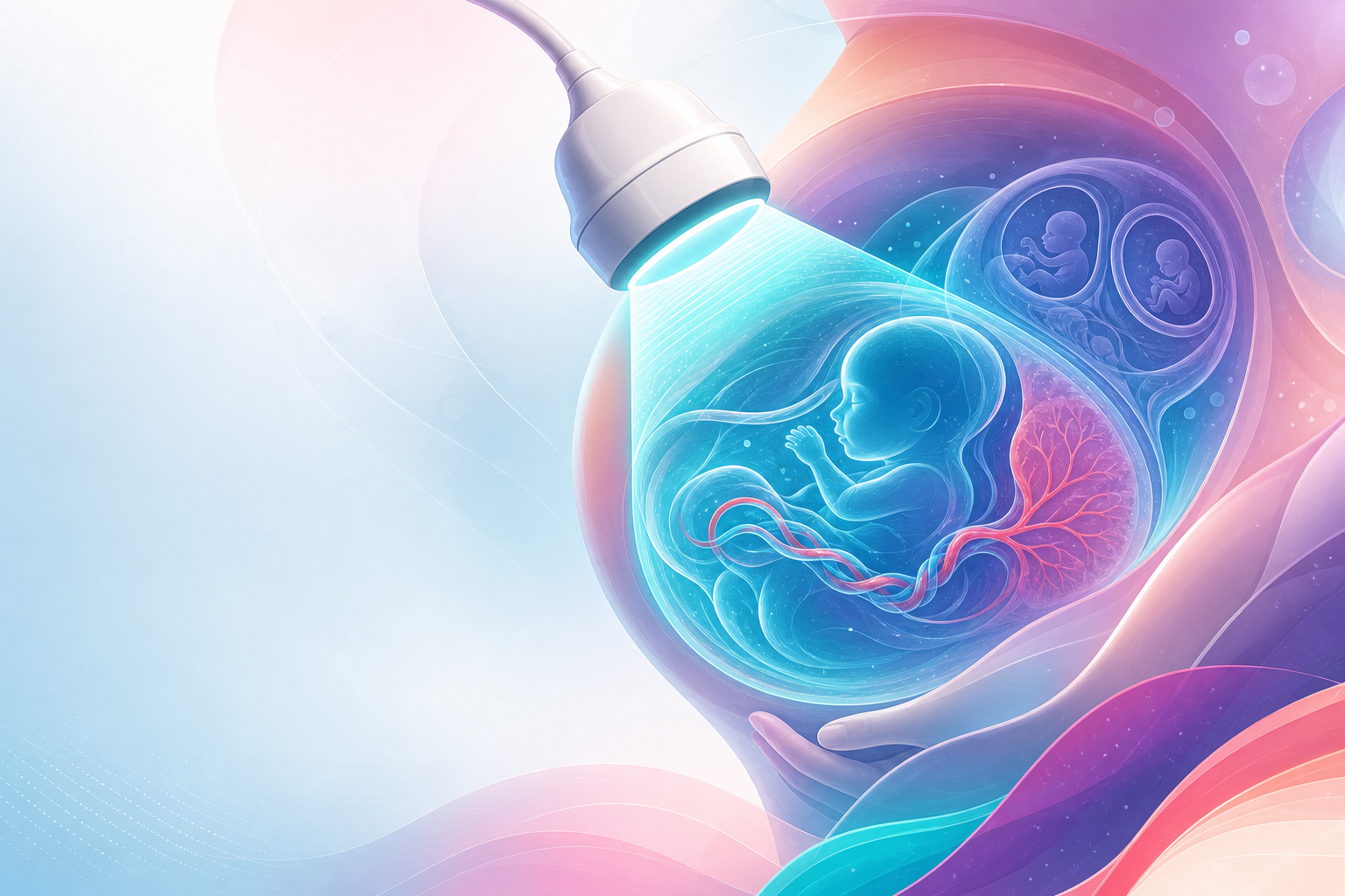
Obstetric ultrasound is the single most important imaging tool in pregnancy and the technical skill the FCOG(SA) examiners expect every registrar to wield safely and interpret correctly. It is not one investigation but a sequence of trimester-specific examinations, each answering a defined clinical question: Is the pregnancy intrauterine, viable and correctly dated? Is the fetus structurally normal and the chromosomal risk acceptable? Is the fetus growing, well-oxygenated and surrounded by normal liquor? Is the placenta normally sited? And — in twins — what is the chorionicity, the single most prognostically important determination in multiple pregnancy.
In the South African public sector ultrasound is a rationed resource. The National Integrated Maternal and Perinatal Care Guideline (NDoH, 2024) builds antenatal care around a basic dating/booking scan and a structured second-trimester anomaly scan where capacity allows, with growth and Doppler surveillance reserved for high-risk pregnancies. Many district hospitals have a single machine and few accredited sonographers; the registrar must therefore know which scan adds clinical value and which is a luxury. This chapter teaches you to use ultrasound — to choose the right examination for the trimester, to perform liquor, placental and Doppler assessment, and to assess multiple pregnancy — rather than merely to list its features. The companion physics and safety material lives in ultrasound-knobology-doppler-safety, and the adnexal-mass interpretation in ultrasound-malignancy-signs.
Core knowledge
Figure F14.1 — Obstetric ultrasound roadmap: ask the right question for the trimester — 1st (location/viability/dating/NT), 2nd (anomaly survey + biometry), 3rd (growth + Doppler) — and scan safely (ALARA; no routine pulsed Doppler in the first trimester).
Safety and the trimester framework
Ultrasound is non-ionising but not without bioeffect: tissue heating (thermal index, TI) and cavitation (mechanical index, MI). ISUOG safety guidance applies the ALARA principle — as low as reasonably achievable — and is most relevant to Doppler, which deposits far more energy than B-mode. Pulsed/spectral Doppler should be avoided in the first trimester for routine indications because the embryo is small and thermally vulnerable; keep TI < 1.0 and exposure time short when Doppler is genuinely indicated. This safety frame is examined repeatedly and detailed in ultrasound-knobology-doppler-safety.
Each trimester has a characteristic task list:
- First trimester (up to ~13⁺⁶ weeks): confirm intrauterine location, number, viability, accurate dating, and chorionicity in twins; offer aneuploidy screening.
- Second trimester (~18–22 weeks): detailed anatomical (anomaly) survey, placental localisation, liquor and early growth.
- Third trimester: growth (fetal biometry and estimated fetal weight), liquor volume, placental site confirmation and, where indicated, fetal Doppler.
Dating and biometry
Crown-rump length (CRL) in the first trimester is the most accurate dating measurement (classically accurate to within ~5 days when measured between roughly 7 and 13 weeks; standard teaching). Once CRL exceeds the validated range, dating shifts to a composite of biparietal diameter (BPD), head circumference (HC), abdominal circumference (AC) and femur length (FL). Dating becomes progressively less reliable with advancing gestation; a scan after ~22 weeks may carry an error of two weeks or more, which is why early dating is prioritised. Dating principles in depth are covered in gestational-age-assessment.
Estimated fetal weight (EFW) is derived from HC, AC and FL (commonly the Hadlock formula; standard teaching), with AC the most sensitive single parameter for growth disturbance because it reflects hepatic glycogen and subcutaneous fat. Serial biometry — not a single measurement — defines growth velocity.
Liquor volume
Amniotic fluid reflects fetal urine output (and therefore renal and placental function) and fetal swallowing. Two semi-quantitative methods are used:
- Deepest vertical pool (DVP, also single deepest pocket): a vertical fluid column free of cord and fetal parts. Standard teaching: normal ~2–8 cm; oligohydramnios < 2 cm; polyhydramnios > 8 cm.
- Amniotic fluid index (AFI): the sum of the deepest pool in each of four uterine quadrants. Standard teaching: normal ~5–25 cm; oligohydramnios < 5 cm; polyhydramnios > 25 cm (mild/moderate/severe bands are used clinically).
Evidence (and current obstetric teaching) favours DVP over AFI for surveillance because AFI diagnoses more oligohydramnios without improving outcomes, leading to more intervention. Liquor pathology — its causes and significance — is the subject of liquor-volume-abnormalities.
Placenta
The anomaly scan localises the placenta relative to the internal os. A placenta whose edge lies within ~2 cm of, or covers, the internal os is low-lying / praevia and mandates a follow-up transvaginal scan in the third trimester, because most low-lying placentas at 20 weeks "migrate" with lower-segment growth. Where there is a previous caesarean scar and an anterior low/praevia placenta, actively look for placenta accreta spectrum (PAS) — loss of the retroplacental clear zone, placental lacunae ("moth-eaten" hypoechoic spaces), bladder-wall interface interruption and increased subplacental vascularity. PAS and praevia carry major haemorrhage risk and link directly to antepartum-haemorrhage.
Fetal Doppler
Doppler interrogates feto-placental and fetal haemodynamics, primarily through resistance indices (pulsatility index, PI; resistance index, RI):
- Umbilical artery (UA): reflects placental vascular resistance. Rising PI, then absent end-diastolic flow (AEDV), then reversed end-diastolic flow (REDV) mark worsening placental insufficiency and are central to growth surveillance.
- Middle cerebral artery (MCA): falling PI signals brain-sparing redistribution. The cerebroplacental ratio (CPR = MCA PI / UA PI), when low, indicates redistribution even when individual vessels are borderline. MCA peak systolic velocity (MCA-PSV) is the non-invasive marker of fetal anaemia (raised in anaemia) — pivotal in rh-isoimmunisation.
- Ductus venosus (DV): a late, ominous sign in early-onset FGR — absent or reversed a-wave reflects cardiac decompensation and informs delivery timing.
These vessels and the underlying physiology of redistribution are developed in placental-insufficiency-response.
Chorionicity in multiples
In twins, chorionicity (number of placentas/outer membranes) matters more than zygosity because monochorionic twins share a placental circulation and risk twin-to-twin transfusion syndrome (TTTS), selective growth restriction and twin anaemia-polycythaemia sequence. Chorionicity is determined most reliably in the first trimester:
- Lambda (λ) / "twin-peak" sign + thick dividing membrane = dichorionic.
- T-sign (membrane meets placenta at right angles, no wedge of placental tissue) + thin membrane = monochorionic.
- Two separate placentas or discordant fetal sex = dichorionic.
Document chorionicity and amnionicity and label twins consistently. Detailed management of multiples is in multiple-pregnancy.
Assessment

First-trimester scan
Begin transabdominally; use the transvaginal probe for early or uncertain findings (better resolution, lower energy to a deeper target). Systematically establish:
- Location — intrauterine gestation sac with a yolk sac then fetal pole; exclude ectopic if the uterus is empty with a positive βhCG.
- Viability — cardiac activity is expected once CRL is ≥ ~7 mm (standard teaching); a mean sac diameter ≥ ~25 mm without a fetal pole, or CRL ≥ ~7 mm without cardiac activity, suggests early pregnancy loss — repeat/second-opinion before declaring non-viability.
- Number and chorionicity — count sacs, fetal poles and yolk sacs; assign chorionicity now (see above).
- Dating — CRL.
- Aneuploidy screening — nuchal translucency (NT) at ~11–13⁺⁶ weeks combined with maternal age, βhCG and PAPP-A; cell-free DNA/NIPT where available. Screening pathways link to antenatal-screening and counselling to down-syndrome-counselling.
Second-trimester anomaly scan (~18–22 weeks)
A structured survey — head and brain (ventricles, posterior fossa, cavum septi pellucidi), face and lips, spine in three planes, the four-chamber heart and outflow tracts, diaphragm, stomach, kidneys and bladder, abdominal wall and cord insertion, and all long bones. Confirm placental site, measure liquor and record biometry. This is the highest-yield single examination for structural anomaly detection; in the SA public sector it is the scan to protect when capacity is limited.
Growth, liquor and Doppler surveillance (third trimester)
Triggered by risk factors — hypertension/pre-eclampsia, previous FGR or stillbirth, decreased-fetal-movements, antepartum haemorrhage, diabetes, or a fundal height lagging dates. Assessment combines:
- Serial biometry / EFW plotted on a growth chart (customised where available); a fetus < 10th centile is small-for-gestational-age — distinguishing constitutionally small from truly growth-restricted is the clinical art, and is the focus of intrauterine-growth-restriction.
- Liquor by DVP.
- UA Doppler as first-line; escalate to MCA/CPR and DV by FGR phenotype and gestation.
Twin surveillance
Dichorionic twins: growth and liquor surveillance, typically less intensive. Monochorionic twins require fortnightly scans from ~16 weeks for TTTS (the classic stuck-twin/polyhydramnios-oligohydramnios sequence with discordant bladders) and growth discordance — link to multiple-pregnancy.
Management

Ultrasound rarely treats; it directs management. The registrar's task is to convert each finding into a clear action and a referral level appropriate to the SA tiered system.
Acting on dating
Set the EDD from the earliest accurate scan (CRL preferred) and do not revise it on later biometry unless the discrepancy is large and the early date was unreliable. Accurate dating underpins screening windows, growth-chart interpretation and decisions about prematurity and prolonged pregnancy. Tie booking and dating logistics back to antenatal-booking.
Acting on liquor
- Oligohydramnios: confirm membranes are intact, exclude FGR and renal/urinary tract anomaly; near term it may prompt delivery, remote from term it prompts surveillance and a search for cause.
- Polyhydramnios: screen for maternal diabetes and fetal anomalies impairing swallowing (e.g. oesophageal atresia, neuromuscular conditions); manage the underlying cause. Full work-up in liquor-volume-abnormalities.
Acting on placental findings
A low-lying placenta at 20 weeks → transvaginal rescan in the third trimester. Confirmed praevia or PAS → refer to a regional/tertiary unit with blood-bank, interventional and surgical capacity; plan a delivery date, antenatal corticosteroids and a senior-led caesarean. Never perform a digital vaginal examination in suspected praevia. This is an antepartum-haemorrhage pathway.
Acting on Doppler — the FGR delivery framework
Doppler primarily informs timing of delivery in growth restriction, balancing stillbirth risk against prematurity:
- Normal UA Doppler with SGA → continued surveillance.
- Raised UA PI / low CPR → increased surveillance, plan steroids, deliver by late preterm/term depending on phenotype.
- AEDV → admit, corticosteroids, intensive monitoring; deliver in the early-preterm window per protocol.
- REDV or abnormal DV (absent/reversed a-wave) → delivery is usually indicated, by caesarean as these fetuses tolerate labour poorly. Synthesise with placental-insufficiency-response and intrauterine-growth-restriction.
MCA-PSV above the threshold (classically > 1.5 multiples of the median; standard teaching) in an at-risk fetus signals significant anaemia → refer for fetal medicine assessment and possible intrauterine transfusion (rh-isoimmunisation).
Acting on chorionicity and TTTS
Monochorionic diamniotic twins need a defined surveillance pathway and a low threshold for referral. Suspected TTTS (Quintero staging) is a fetal-medicine emergency — refer urgently to a tertiary unit offering fetoscopic laser; delay loses fetuses. Detailed staging and intervention are in multiple-pregnancy.
Emergency drill — the empty uterus with a positive pregnancy test
DRILL — suspected ruptured ectopic. A haemodynamically unstable woman with a positive βhCG and an empty uterus on scan (± free fluid in the pouch of Douglas/Morison's pouch) is a ruptured ectopic until proven otherwise.
- Call for help; resuscitate — two large-bore IV lines, bloods including cross-match, fluids.
- Do NOT wait for serial βhCG in the shocked patient.
- Theatre immediately for laparotomy/laparoscopy — do not delay for imaging perfection. Hand off ongoing management per the ectopic pathway. Free fluid + empty uterus + instability = operate.
SA level-of-care and resource framing
- District (level 1): dating/booking scan, viability, gross placental site, basic liquor — sufficient to triage and date most pregnancies. Many sites rely on a midwife/medical officer with focused training.
- Regional (level 2): anomaly scanning, growth and UA Doppler surveillance, twin follow-up.
- Tertiary/fetal medicine (level 3): advanced Doppler, intrauterine transfusion, fetoscopic laser for TTTS, PAS surgery.
The 5th-edition Maternity Guideline (2024) reserves intensive Doppler and serial growth surveillance for defined high-risk indications precisely because machine and sonographer access is limited; the registrar adds most value by selecting and referring appropriately, and by ensuring HIV-positive women — a large share of the SA antenatal population — receive the same structured dating, anomaly and growth pathway, with no contraindication to ultrasound itself.
Red flags / pitfalls
- Declaring non-viability too early or on one scan. Respect the CRL/MSD thresholds, repeat after an interval, and obtain a second opinion. A wrong call can end a wanted, viable pregnancy.
- Missing the ectopic. An empty uterus with a positive βhCG is ectopic until excluded — never reassure on a "normal pelvic scan" without confirming an intrauterine pregnancy.
- Assigning chorionicity late. After the first trimester chorionicity is far harder to call; a missed monochorionic diagnosis means missed TTTS surveillance.
- Pulsed Doppler in the first trimester for routine indications — an avoidable ALARA breach; B-mode confirms viability.
- Over-diagnosing oligohydramnios with AFI and intervening unnecessarily — prefer DVP for surveillance.
- Revising a reliable early EDD on late biometry — a small fetus is then mislabelled "wrong dates" and FGR is missed.
- Digital examination in suspected praevia — can precipitate catastrophic haemorrhage.
- Treating SGA and FGR as identical — a constitutionally small, well-Dopplered fetus does not warrant the same intervention as one with AEDV/REDV.
- Over-reading PAS — or under-reading it. In any anterior low/praevia placenta over a caesarean scar, deliberately look for accreta and refer; failing to plan is a leading avoidable cause of massive obstetric haemorrhage.
- Forgetting the operator-dependence of the modality. Image quality, completeness and honest documentation of suboptimal views are part of the standard of care.
Evidence anchors
- SA National Integrated Maternal and Perinatal Care Guideline (NDoH, 2024) — the South African source of truth: antenatal scan schedule, dating, indications for growth/Doppler surveillance, and the tiered (district/regional/tertiary) referral framework within which obstetric ultrasound is rationed.
- ISUOG ultrasound safety guidance — the ALARA principle and thermal/mechanical index limits; caution with pulsed Doppler in the first trimester (see ultrasound-knobology-doppler-safety).
- RCOG Green-top Guideline No. 31 — Small-for-Gestational-Age and Growth-Restricted Fetus, with ISUOG Doppler standards (umbilical and middle cerebral artery, ductus venosus, cerebroplacental ratio) — the basis of the FGR surveillance and delivery-timing framework.
- RCOG Green-top Guideline No. 51 — Management of Monochorionic Twin Pregnancy — chorionicity-based surveillance, TTTS and the monochorionic scanning schedule.
- RCOG Green-top Guideline No. 27a/27 (Placenta Praevia and Placenta Accreta) — ultrasound diagnosis of praevia and the placenta accreta spectrum and the referral/delivery pathway.
- RCOG Green-top Guideline No. 57 — Reduced Fetal Movements — a common trigger for growth/liquor/Doppler assessment.
- NICE NG201 — Antenatal care (2021) — dating-scan and anomaly-scan principles, cross-referenced where SA practice aligns.
- NICE TA156 (anti-D) with MCA-PSV as the non-invasive marker of fetal anaemia in red-cell alloimmunisation (see rh-isoimmunisation).
- Hedged as standard teaching, not line-itemed in verified sources: CRL dating accuracy (~±5 days), DVP/AFI numeric thresholds (DVP 2–8 cm; AFI 5–25 cm), Hadlock EFW formula, viability cut-offs (CRL ≥7 mm, MSD ≥25 mm), and MCA-PSV > 1.5 MoM. These are conventional ultrasound thresholds; confirm exact local protocol values before clinical use.
