Clinical overview
Minimally invasive surgery (MIS) — laparoscopy and hysteroscopy — has displaced laparotomy for most benign gynaecological operations because it reduces blood loss, pain, wound complications, adhesion formation and length of stay. But the trade-off is a distinctive, often iatrogenic complication profile that is largely preventable and largely front-loaded: most serious laparoscopic injuries happen at entry, before the operation has even begun, and most hysteroscopic disasters relate to the distension medium rather than the surgical target. The registrar examined on this objective is being asked to describe methods of preventing complications — so the emphasis is squarely on the systems, techniques and checks that stop harm, not merely on listing the harms.
The framing that earns marks is prevention across the whole operative pathway: patient selection and preoperative optimisation; safe entry; energy and instrument discipline; recognition of vascular, visceral and urological injury; control of the distension/insufflation environment; and structured recovery. In the South African public setting these principles must be read against real constraints — variable laparoscopic stack availability, theatre lists dominated by emergency obstetrics, a high background of pelvic adhesive disease from sexually transmitted infection and prior caesarean, and a substantial HIV-positive surgical population. Prevention here is not abstract: a single recognised-and-repaired bowel injury is the difference between a one-week recovery and a death from delayed peritonitis. This chapter builds on the related technical objectives — electrosurgery-safety, pneumoperitoneum, operative-hysteroscopy, surgical-instruments-safe-use and eras-principles — and should be read alongside them.
Core knowledge
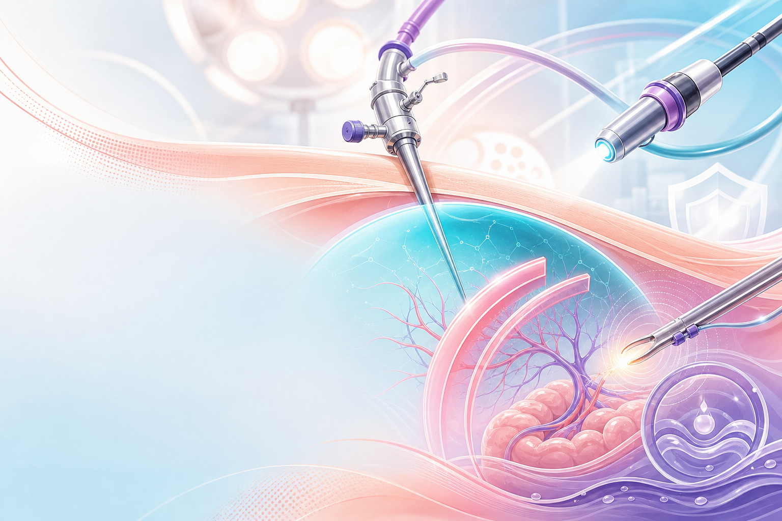
Figure F7.1 — Safe laparoscopic entry: risk scan, time-out, choosing the entry (Veress / Hasson / Palmer’s point), the port-placement map and the key entry risks — prevent the injury before the operation starts.
The complication map
Laparoscopic complications cluster predictably, and naming the cluster is half the prevention:
- Entry injuries — the single most dangerous category. Vascular injury (aorta, common iliac vessels, inferior epigastric vessels) and visceral injury (bowel, especially with adhesions to the umbilicus) occur at Veress needle or primary trocar insertion. Major vessel injury is rare but is the leading cause of laparoscopy-related death.
- Visceral injury — bowel, bladder, ureter. Thermal injury from electrosurgery is particularly dangerous because it presents late (typically 3–10 days postoperatively as delayed perforation), having looked benign at the time.
- Urological injury — bladder injury during dissection of the bladder flap or anterior compartment; ureteric injury at the pelvic side wall, infundibulopelvic ligament, uterine artery and uterosacral ligaments — the four classic danger zones.
- Insufflation-related — CO₂ embolism, subcutaneous emphysema, pneumothorax/pneumomediastinum, and the cardiorespiratory consequences of raised intra-abdominal pressure and steep Trendelenburg.
- Port-site complications — bleeding from the inferior epigastric vessels, port-site hernia (risk rises with fascial defects ≥10 mm), and port-site metastasis in malignancy.
- Hysteroscopic complications — uterine perforation, distension-fluid overload with dilutional hyponatraemia (the hysteroscopic equivalent of TURP syndrome), gas embolism, false passage, cervical trauma and infection.
Entry physiology and anatomy
The umbilicus is the standard entry point because the abdominal wall layers fuse there, but it sits only a few centimetres anterior to the aortic bifurcation and great vessels in a thin patient — hence the insistence on a 45° insertion angle and awareness of body habitus. Insufflation to an adequate pneumoperitoneum lifts the anterior wall away from the viscera and great vessels, which is why entry safety is intimately tied to confirming correct Veress placement before gas is delivered. The inferior epigastric vessels run lateral to the obliterated umbilical arteries and medial to the round ligament — lateral ports must be placed under direct vision and lateral to these vessels (transilluminating the wall helps in slim patients but is unreliable when subcutaneous fat is thick). See pneumoperitoneum for the physiology in detail.
Energy and the late-injury problem
Electrosurgical injury prevention depends on understanding why thermal damage is insidious: lateral thermal spread, capacitive coupling, insulation failure and direct coupling can all deliver energy to bowel that is not in the field of view. The injured serosa may look intact, devitalise over days, then perforate. This is the rationale for keeping all active electrode tips in view, inspecting instrument insulation, using the lowest effective power, favouring bipolar or advanced vessel-sealing devices for pedicles, and being cautious with monopolar near bowel and ureter. The detail belongs to electrosurgery-safety, but the prevention principle is examinable here: a thermal bowel injury you cannot see is the one that kills your patient a week later.
Assessment
Preoperative selection and optimisation
Prevention starts before theatre. A structured preoperative assessment — covered fully in preoperative-performance-status — should establish functional capacity (METs), the ASA physical-status class, and the specific MIS risk factors:
- Adhesion risk: previous laparotomy, midline scar, prior peritonitis, severe endometriosis, pelvic inflammatory disease. A periumbilical scar or suspected bowel adherence to the umbilicus is a direct indication for an alternative entry site (left upper quadrant / Palmer's point) rather than blind umbilical entry.
- Body habitus: both very low and very high BMI distort the umbilicus–vessel relationship and change the safe entry angle.
- Cardiorespiratory reserve: raised intra-abdominal pressure and Trendelenburg reduce venous return and lung compliance — relevant in cardiac disease and severe respiratory disease.
- Anticoagulation, anaemia, and VTE risk: optimise haemoglobin where feasible, plan thromboprophylaxis.
- HIV status and immune function: in the SA population, document HIV status and ART (TLD — tenofovir/lamivudine/dolutegravir — per the SA ART guidelines); immunosuppression and the higher background of pelvic infection/adhesions raise the threshold for difficulty. HIV status does not contraindicate MIS, and universal precautions apply to every case regardless of serostatus.
Intra-operative vigilance and recognition
The assessment that prevents a minor injury becoming a fatal one is intra-operative recognition:
- After primary trocar insertion, perform an immediate 360° panoramic inspection of the entry site and great vessels for bleeding or visceral injury before proceeding.
- Watch the insufflation pressure–volume relationship at entry: low opening pressure (classically <8–10 mmHg before flow rises) supports correct intraperitoneal Veress placement, while high opening pressure suggests preperitoneal or visceral placement (standard teaching, not a single-trial threshold).
- At the end of every laparoscopy, inspect all port sites under vision while withdrawing trocars to catch abdominal-wall bleeding, and re-survey the pelvis for haemostasis at reduced insufflation pressure (bleeding tamponaded by 15 mmHg of CO₂ can reopen on deflation).
- In hysteroscopy, continuous monitoring of fluid balance (inflow minus outflow) is the central assessment that prevents fluid-overload catastrophe — assign a dedicated person to track the deficit in real time.
Postoperative surveillance for occult injury
Because the worst MIS injuries declare late, postoperative assessment is a prevention tool. Pain, tachycardia, fever, ileus or peritonism beyond the expected mild postoperative course must be treated as a missed bowel injury until proven otherwise — not as "slow recovery" or "constipation". A patient who is more unwell on day 3 than on day 1 after laparoscopy needs active exclusion of visceral injury, not reassurance and discharge.
Management

Management of this objective means the preventive manoeuvres and the response when a complication is recognised. Work through the operative pathway.
1. Team and checklist discipline
Every case begins with the WHO Surgical Safety Checklist — sign-in, time-out, sign-out — confirming patient, procedure, consent (see informed-consent), allergies, antibiotic prophylaxis, VTE prophylaxis, equipment availability and anticipated blood loss. The time-out is the moment to confirm the entry plan, the energy devices in use, and the contingency for conversion to laparotomy. Equipment is checked before induction: insufflator and tubing, light source, camera white-balance, suction-irrigation, and insulation integrity of every reusable electrosurgical instrument.
2. Safe entry
There is no single proven-superior entry technique for all patients; the evidence (synthesised in RCOG Green-top Guideline 49, Preventing Entry-related Gynaecological Laparoscopic Injuries) supports a disciplined technique rather than one dogma. Core preventive measures:
- Empty the bladder (catheterise) and consider gastric decompression to lower bladder and stomach injury risk.
- Veress (closed) entry: insert at the base of the umbilicus at the correct angle for habitus; confirm intraperitoneal placement (low opening pressure; the various "safety tests" are confirmatory, not definitive); insufflate to an adequate pneumoperitoneum before inserting the primary trocar. Insufflating to a higher intra-abdominal pressure (often quoted ~20–25 mmHg) transiently for trocar insertion only increases the distance to the great vessels and the splinting of the wall — drop the pressure to a working level once ports are in.
- Open (Hasson) entry: a reasonable alternative, particularly where adhesions are anticipated; it does not eliminate bowel injury but allows it to be recognised immediately.
- Alternative entry (Palmer's point, left upper quadrant) when periumbilical adhesions, prior midline surgery or failed umbilical entry are present — first exclude splenomegaly and decompress the stomach.
- Secondary ports under direct vision, placed lateral to the inferior epigastric vessels.
3. Energy and dissection discipline
Apply the electrosurgery-safety principles: lowest effective power, active electrode always in view, prefer bipolar/advanced bipolar or ultrasonic devices for vascular pedicles, inspect insulation, avoid open activation and capacitive coupling. Identify and protect the ureter before dividing the infundibulopelvic ligament, uterine artery and uterosacral ligaments — restore anatomy and skeletonise rather than dividing blind tissue. Maintain meticulous haemostasis; do not rely on pneumoperitoneum to mask a bleeding pedicle.
4. Recognising and managing injury — the emergency drills
MAJOR VASCULAR INJURY AT ENTRY — this is a life-threatening emergency. Drill:
- Do NOT remove the offending instrument — leave the trocar/Veress in situ to tamponade.
- Call for help immediately — senior surgeon, vascular surgeon if available, alert anaesthetist and theatre team out loud.
- Convert to laparotomy without delay for direct compression and repair.
- Activate massive transfusion — large-bore IV access, cross-match/group-specific or emergency O-negative blood, warmed fluids, tranexamic acid 1 g IV for major surgical haemorrhage.
- Apply direct pressure on the vessel while definitive control is achieved.
BOWEL INJURY: if recognised intra-operatively, repair (primary repair or resection per the injury) — get general/colorectal surgery input; a small sharp serosal injury may be over-sewn, but thermal injury requires resection of the surrounding devitalised zone because the true injury extends beyond the visible burn. If suspected postoperatively (day-3 unwell patient), resuscitate, image (CT), give broad-spectrum antibiotics and take back to theatre — never observe a deteriorating post-laparoscopy patient.
BLADDER INJURY: recognised cystotomy is repaired in layers with prolonged catheter drainage; small injuries may be managed by catheter drainage alone. URETERIC INJURY: the danger is the unrecognised injury — if in doubt, cystoscopy ± retrograde study; manage with stenting or reimplantation per urology and the level of expertise available.
5. Insufflation and positioning safety
Use the lowest working intra-abdominal pressure that gives adequate exposure; communicate with anaesthesia about Trendelenburg tolerance; secure the patient against sliding; protect pressure points and brachial plexus. CO₂ embolism (sudden hypotension, hypoxia, a "mill-wheel" murmur, fall in end-tidal CO₂) is an anaesthetic emergency: stop insufflation, deflate, place left-lateral head-down (Durant) position, give 100% oxygen, and support the circulation.
6. Hysteroscopy-specific prevention
For operative-hysteroscopy, the dominant preventable disasters are perforation and fluid overload:
- Distension media discipline: where monopolar resection requires non-electrolyte media (e.g. glycine/sorbitol), dilutional hyponatraemia and fluid overload are the lethal risk — strict, continuously monitored fluid-deficit limits with automated fluid-management systems. Bipolar systems use normal saline, which removes the hyponatraemia risk but not the volume-overload risk. The AAGL hysteroscopy fluid-management guidance frames these deficit thresholds (verify the exact litre limits against the current AAGL document before quoting — standard teaching uses lower limits for hypotonic non-electrolyte media than for isotonic saline).
- Stop and reassess at the deficit limit, treat overload with fluid restriction and diuresis, check sodium urgently, and beware acute hyponatraemic cerebral oedema.
- Perforation: minimise by adequate cervical preparation/dilatation, gentle technique under vision, and never advancing energy or instruments blindly; a perforation with an energy device or with possible bowel involvement mandates laparoscopy to exclude visceral injury.
- Air/gas embolism: avoid the Trendelenburg-with-open-cervix pump effect; flush tubing of air.
7. ERAS and recovery
Apply eras-principles — early mobilisation, early feeding, multimodal opioid-sparing analgesia, judicious peri-operative fluids (perioperative-fluids), early catheter removal, and VTE prophylaxis — to reduce the medical complications (ileus, VTE, chest infection) that compound the surgical ones.
South African context
In SA practice, prevention is shaped by resource reality. Laparoscopic capacity is concentrated at regional and tertiary levels; many district hospitals operate by laparotomy, so part of "prevention" is appropriate referral and case selection — not attempting advanced MIS without the stack, the support and the conversion capability. The National Department of Health (NDoH) National Integrated Maternal and Perinatal Care Guideline (NDoH, 2024) and the SA levels-of-care framework define which facility should undertake which procedure; the Saving Mothers / NCCEMD reports repeatedly identify delayed recognition and operating beyond a facility's capability as avoidable contributors to death — a lesson that transfers directly to gynaecological MIS. Universal precautions and safe sharps handling matter in a high-HIV population; emergency blood follows the institutional protocol and the SA EML. The high background of post-PID and post-caesarean pelvic adhesions raises entry risk, lowering the threshold for open or alternative-site entry.
Red flags / pitfalls

- Blind faith in "the safety tests." No single Veress confirmation test is definitive; opening pressure is the most reliable single sign, but technique and anatomy awareness matter more than any one test.
- Missing the late thermal bowel injury. The classic exam (and real-life) trap: discharging a day-1-well patient who is unwell on day 3. Tachycardia + pain + fever after laparoscopy is a visceral injury until excluded — do not relabel it constipation or "slow recovery".
- Leaving the trocar out in major vascular injury. Removing the tamponading instrument before laparotomy turns a controllable injury into exsanguination. Leave it in, get help, open.
- Deflating without re-checking haemostasis. A pedicle compressed by 15 mmHg of CO₂ can bleed once the gas is out — always re-survey at low pressure and inspect ports on withdrawal.
- Ignoring the hysteroscopic fluid deficit. Treat the deficit number as a hard stop, not a guideline to "keep an eye on". Hyponatraemic cerebral oedema and pulmonary oedema are preventable by stopping in time.
- Operating beyond facility capability. Attempting advanced MIS without a stack, blood, conversion capacity or senior support — an avoidable-factor theme in Saving Mothers thinking.
- Lateral ports placed blind. The inferior epigastric vessels are injured by ports sited medially or placed without direct vision; transillumination is unreliable in obese patients.
- Skipping the checklist or the time-out because the case "feels routine". The WHO checklist exists precisely to catch the routine case that goes wrong.
Evidence anchors
- RCOG Green-top Guideline No. 49 — Preventing Entry-related Gynaecological Laparoscopic Injuries — the primary reference for safe entry technique, alternative entry sites, and recognition/management of entry injuries.
- WHO Surgical Safety Checklist — sign-in/time-out/sign-out; the systems-level prevention backbone.
- AAGL guidance on electrosurgery and on hysteroscopic distension-media fluid management — energy safety principles and fluid-deficit limits for hysteroscopy (verify current AAGL deficit thresholds before quoting exact figures).
- ERAS Society gynaecologic/oncology guidelines — perioperative recovery measures that reduce medical complications (see eras-principles).
- WOMAN trial (Lancet 2017) — tranexamic acid 1 g IV early in major obstetric haemorrhage; the principle of early TXA in major surgical bleeding informs the vascular-injury drill.
- SA NDoH National Integrated Maternal and Perinatal Care Guideline (NDoH, 2024) and Saving Mothers / NCCEMD reports — SA levels-of-care, referral discipline, and the recurring avoidable-factor lessons (delayed recognition; operating beyond capability).
- SA HIV/ART Consolidated Guidelines (2023) and SA EML (Hospital Level) — context for the HIV-positive surgical patient and emergency blood/medication availability.
Note on hedged facts: the Veress opening-pressure cut-off (~8–10 mmHg), the high-pressure entry value (~20–25 mmHg for trocar insertion only), the delayed thermal-injury window (~3–10 days), the ≥10 mm port-site-hernia threshold, and the hysteroscopic fluid-deficit limits are stated as standard surgical teaching and are deliberately not pinned to a single trial — verify exact thresholds against the current RCOG GTG 49 and AAGL documents before quoting numerically in an exam answer.
