Clinical overview
Electrosurgery is the most ubiquitous energy modality in the gynaecological theatre, used in almost every laparoscopy, hysteroscopy, laparotomy and vaginal procedure for cutting and haemostasis. Precisely because it is so routine, its hazards are under-appreciated: the same physics that lets us coagulate a pedicle can perforate a bowel out of the surgeon's field of view, burn the patient at a distant skin site, ignite an airway or alcohol-prep fire, or interfere with a cardiac implantable electronic device. Energy-related injury — particularly bowel and ureteric thermal injury at laparoscopy — is a recurrent theme in medicolegal claims and in surgical morbidity audits, and a substantial proportion of these injuries are not recognised at the time of operation. The registrar who understands why electrosurgery behaves as it does will avoid the errors that the surgeon who simply "presses the yellow pedal" cannot.
This is a "discuss issues related to electrical safety" objective, so the weight sits in Core knowledge (the physics and the failure mechanisms) and Management (the safe-use drills and what to do when something goes wrong). The discussion must be genuinely mechanistic: examiners expect you to explain capacitive coupling, the difference between cut and coagulation waveforms, why a "no return-electrode" alarm is a patient-safety event, and how monopolar and bipolar circuits differ — not merely to recite a list of rules. In the South African context the issues are sharpened by the mix of older and donated electrosurgical units in district and regional theatres, variable biomedical-engineering maintenance, the high prevalence of HIV (sharps and plume exposure matter for the whole team), and the reality that the registrar is often the most senior person in a level-1 or level-2 theatre at night. This chapter sits alongside surgical-instruments-safe-use, mis-complication-prevention, pneumoperitoneum, operative-hysteroscopy and eras-principles.
Core knowledge
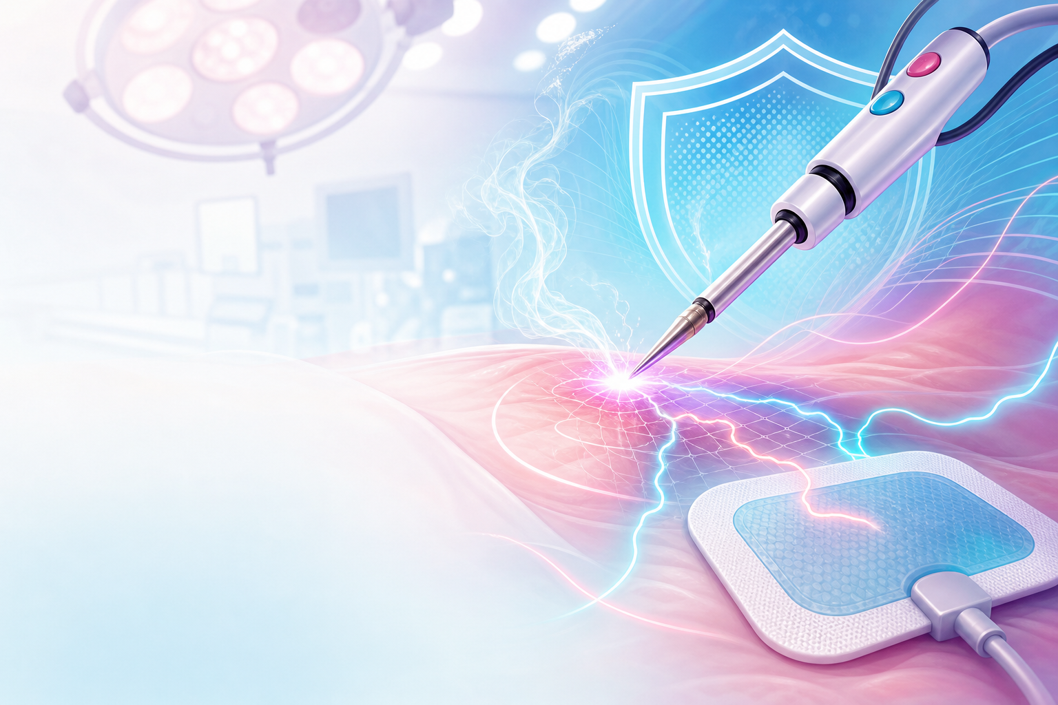
Figure F4.1 — Electrosurgery is current, not cautery: the patient is part of the circuit, power density (small tip vs large pad) decides the tissue effect, waveform decides cut versus coagulation, and bipolar confines current to the tissue between the jaws.
The basic circuit and what electrosurgery actually does
Electrosurgery uses high-frequency alternating current (radiofrequency, classically in the hundreds of kilohertz to low megahertz range) to generate heat within tissue. The key conceptual point is that the patient is part of an electrical circuit: current flows from the generator, through the active electrode, through the patient's tissue, and back to the generator. Tissue has electrical resistance (impedance), and current passing through a resistance generates heat — this is the thermal effect that cuts and coagulates. The reason high frequency is used is the frequency-response of excitable tissue: ordinary low-frequency mains current (50 Hz in South Africa) depolarises nerve and cardiac muscle and causes electrocution, whereas radiofrequency current oscillates far too fast to depolarise membranes, so it produces heat without neuromuscular stimulation.
Do not confuse electrosurgery ("diathermy") with electrocautery. In true electrocautery a current heats a wire, and it is the hot wire that touches and burns the tissue — current never enters the patient. In electrosurgery the current itself passes through the patient and generates heat in the tissue. Almost everything in a modern gynaecology theatre (the "Bovie", LigaSure, bipolar forceps, the hysteroscopic resectoscope) is electrosurgery, not cautery.
The tissue effect depends on power density — current concentrated into a small area produces intense, localised heating; the same current spread over a large area barely warms tissue. This single principle explains both how we cut (tiny contact area at the electrode tip → very high power density → rapid vaporisation) and why the return electrode is safe (huge contact area → low power density → negligible warming).
Cut, coagulation and the waveform
The generator shapes the current into different waveforms, and the registrar should be able to explain the difference:
- Cut (pure cut): a continuous (undamped), relatively low-voltage sinusoidal waveform. Continuous energy delivery superheats intracellular water so quickly that cells explode (vaporisation), producing a clean cutting effect with little lateral thermal spread or coagulation.
- Coagulation: an interrupted (damped, "on" only a small fraction of the time), much higher-voltage waveform. The intermittent high-voltage bursts dehydrate and denature tissue rather than vaporising it, producing coagulation/desiccation and, with non-contact sparking, fulguration. The higher peak voltage of coag mode is exactly why it is the mode most associated with capacitive coupling and insulation-failure injuries.
- Blend: intermediate duty cycles giving simultaneous cutting with some haemostasis.
A common teaching trap: "cut" is lower voltage than "coag", and lateral thermal spread is a property of energy delivered over time, not of the mode name alone — prolonged "cutting" near the ureter is still dangerous.
Monopolar versus bipolar
This is the single most examinable distinction.
- In monopolar electrosurgery the active electrode is the instrument tip; current travels through the patient's whole body to a return (dispersive) electrode — the "patient plate" or pad — and back to the generator. The current path is long and partly unpredictable. Monopolar is versatile (cut, coag, fulgurate) but carries the risks of return-electrode burns, alternate-site burns, capacitive coupling and stray-current injury.
- In bipolar electrosurgery the two electrodes are the two jaws of the forceps; current flows only through the small bridge of tissue held between them. There is no return pad and no current through the rest of the patient, so most of the monopolar-specific hazards are abolished. Bipolar is inherently the safer modality and is preferred for coagulating vascular pedicles laparoscopically, near the ureter and bowel, and in patients with cardiac devices. Its limitation is that classic bipolar coagulates but does not cut; advanced bipolar vessel-sealing devices (e.g. LigaSure-type systems) use feedback-controlled energy to seal and then divide vessels and have largely displaced clips and sutures for many pedicles.
The mechanisms of electrosurgical injury
A safe surgeon thinks in terms of how current can reach tissue it should not. The recognised failure modes — emphasised in RCOG guidance on laparoscopic injuries (GTG 49) and in AAGL energy-safety teaching — are:
- Direct application / inadvertent activation: the commonest cause. The pedal is pressed while the active tip touches or is near non-target tissue, or the activated tip is moved out of view. Always activate only when the working tip is in full view and clear of bowel, bladder, vessels and ureter.
- Direct coupling: the activated monopolar electrode touches another conductive instrument (a metal grasper, a laparoscope) which then conducts current to whatever it is touching, often out of view.
- Insulation failure: a breach in the insulating sheath of a monopolar instrument lets current escape at an unseen point along the shaft — frequently outside the laparoscopic field of view, so the burn to bowel is silent. High-voltage coag mode makes a small defect far more dangerous. Inspect instrument insulation before every case.
- Capacitive coupling: a capacitor is formed whenever a conductor (the active electrode) carrying high-frequency current is separated by an insulator from another conductor. Current can be induced across the insulator without any direct contact — for example into a metal cannula, or into the laparoscope. If that induced charge then concentrates onto a small area of tissue (e.g. a hybrid metal-and-plastic port system that traps the charge), a burn results. Capacitive coupling is worsened by high voltage (coag), long activation, narrow electrodes inside cannulae, and mixed metal/plastic port systems. Using all-metal or all-plastic cannula systems, low-voltage cut where possible, short bursts, and active-electrode monitoring (AEM) instruments mitigate it.
- Return-electrode (dispersive pad) burns: if the return pad is poorly applied (small effective contact area, lifting at an edge, placed over scar, bone or hair, or over a metal prosthesis), power density at the pad rises and the patient is burned at the pad site. Modern generators use a split (dual-foil) return electrode with contact-quality monitoring (return-electrode monitoring, REM); the generator measures impedance between the two foils and disables output if contact is inadequate.
Smoke plume, fire and cardiac devices
Surgical plume contains particulate, viable cells, toxic gases and potentially infectious material; intact viral DNA (e.g. HPV) has been recovered from plume, which is a relevant occupational concern in a high-HPV, high-HIV population. The standard mitigation is smoke evacuation and well-fitting respiratory protection. Surgical fires require the classic triad — an oxidiser (oxygen, nitrous oxide), an ignition source (the active electrode), and fuel (alcohol-based skin prep, drapes, bowel gas/methane, hair). Allowing alcohol prep to dry fully and avoiding pooling, and caution with bowel gas during open procedures, are the practical lessons. Cardiac implantable electronic devices (pacemakers, ICDs) can be affected by monopolar current: prefer bipolar, keep the return-pad current path away from the device, use short bursts, and arrange perioperative device management (magnet/programming and monitoring) in liaison with cardiology.
Assessment
"Assessment" in this objective means risk-assessing the patient, the equipment and the procedure before energy is used, and recognising injury intra- and post-operatively.
Pre-operative and equipment assessment
- Patient factors: cardiac implantable device (type, dependence, last check), metal implants/prostheses near a potential current path, body habitus and skin condition affecting pad placement, and prior surgery raising the chance of adhered bowel near operative sites.
- Equipment check (a deliberate routine, not an afterthought): confirm the generator passes its self-test; inspect every monopolar instrument's insulation along its full length for cracks; confirm the return electrode is intact and that the generator's contact-quality monitor is enabled; confirm correct device for the task (bipolar/vessel-sealer for pedicles near ureter/bowel). In SA district and regional theatres, where electrosurgical units may be older or donated, the registrar should know that biomedical-engineering servicing and electrical-safety testing of theatre equipment is a governance requirement, and should not use a unit that has failed safety testing or whose alarms have been "worked around".
- Team brief / WHO Surgical Safety Checklist: energy-device readiness, return-pad placement, smoke evacuation and fire risk (alcohol prep, oxygen) belong in the sign-in/time-out, consistent with the WHO Surgical Safety Checklist and ERAS perioperative-safety principles.
Recognising injury
- Return-electrode / alternate-site burn: an unexplained full-thickness skin burn at the pad site or at an unintended grounded contact point — inspect the pad site at the end of every case.
- Thermal visceral injury (the dangerous, often-delayed one): a monopolar electrothermal bowel injury frequently does not bleed and may look like a small blanched area at operation, or may not be seen at all if it occurred out of view via insulation failure or capacitive coupling. The coagulative necrosis evolves over days, so the patient classically re-presents 3–7 days post-laparoscopy with low-grade fever, increasing localised then generalised pain, ileus, tachycardia and rising inflammatory markers — a presentation that must trigger urgent re-imaging and surgical review for delayed bowel perforation rather than reassurance. Ureteric thermal injury may present with flank pain, fever, urinoma or a rising creatinine.
Management

Safe-use drill (the routine that prevents most injuries)
Make these habitual; in an exam, state them as a deliberate sequence:
- Lowest effective settings. Use the lowest power and voltage that achieves the effect; prefer cut (lower-voltage) waveform over high-voltage coag where a choice exists; deliver short bursts, not prolonged activation, to limit lateral thermal spread.
- Activate only under direct vision. Never press the pedal unless the entire working tip is in view and clear of bowel, bladder, vessels and ureter; never activate against, or in contact with, another metal instrument.
- Park the active electrode safely. Between activations keep the monopolar tip in an insulated holster/quiver, never resting on drapes or the patient — this prevents inadvertent-activation fires and burns.
- Inspect insulation of every monopolar instrument before use; discard or repair breached instruments. Consider active-electrode-monitoring (AEM) systems for laparoscopy, which continuously shield against and detect insulation failure and capacitive coupling.
- Bipolar by default near vital structures. Use bipolar or an advanced vessel-sealing device for pedicles and dissection adjacent to ureter, bladder and bowel; respect a margin from the ureter and consider the thermal-spread zone, not just the visible char.
- Return pad correctly placed: clean, dry, well-vascularised muscle mass (commonly thigh/flank), close to the operative field, away from bony prominences, scar, hair and metal implants, with the long axis oriented toward the field. Never bypass a "poor pad contact" / REM alarm — investigate and re-site the pad; the alarm is a patient-safety event.
- Fire prevention: let alcohol-based prep dry fully and avoid pooling; keep the active electrode away from oxygen-enriched fields; be cautious with bowel gas at laparotomy. Know where the theatre's fire response and the generator's off-switch are.
- Plume control: smoke evacuation and appropriate respiratory protection for the whole team, given the HPV/infectious-particle and HIV occupational context.
When an injury is suspected — emergency drill
STOP THE INJURY. Release the foot pedal / deactivate immediately. Do not reactivate.
If an electrosurgical injury is suspected or seen, manage it as a surgical emergency:
- Recognise and disclose: stop, inspect, and call the senior/consultant — energy injuries recognised and repaired at the index operation do far better than those missed.
- Inspect for the full extent: thermal injury extends beyond the visible char; for a bowel contact injury, plan to assess and, where indicated, resect the devitalised margin rather than oversew a deceptively small white patch. Involve general surgery/urology early for bowel or ureteric injury.
- A burn at the pad or an alternate site is documented, photographed, treated as a thermal burn, and reported as a critical incident/adverse event so the generator and pad are quarantined and checked by biomedical engineering.
- Delayed presentation (the night-shift scenario): a patient re-presenting several days after laparoscopy with worsening pain, fever, tachycardia and ileus has a delayed thermal bowel perforation until proven otherwise — resuscitate, image (CT), give broad-spectrum antibiotics, and escalate for laparotomy/laparoscopy and surgical review. Do not discharge with reassurance.
- Document and report: record waveform/power used, device, suspected mechanism, and complete the institutional adverse-event report; quarantine the implicated equipment for safety testing.
South African context
In district (level 1) and many regional (level 2) theatres the registrar may be operating with older or donated electrosurgical units, variable biomedical-engineering support, and no advanced energy devices. The safe response is conservative: rely on bipolar and conventional ligation/suture for haemostasis where vessel-sealers are unavailable; do not improvise around failed safety alarms; and have a low threshold to refer or transfer complex energy-dependent surgery (and any suspected major visceral energy injury) to a centre with general surgery, urology and critical-care backup, consistent with the levels-of-care referral framework in the NDoH service-platform structure. Occupational sharps and plume exposure carry weight in a high-HIV setting — apply standard precautions and post-exposure protocols for the whole team.
Red flags / pitfalls

- The silent out-of-view burn. The most dangerous monopolar injuries (insulation failure, capacitive coupling, direct coupling) happen outside the laparoscopic field of view and are not seen at operation. If you only watch the tip, you will miss them — inspect insulation, use AEM, and keep a high index of suspicion post-operatively.
- "It looked like a tiny white mark." Electrothermal bowel injury looks trivial and bleeds little; the necrosis evolves over days. Underestimating the margin of devitalised tissue (oversewing rather than resecting) is a classic cause of subsequent perforation.
- Bypassing the return-electrode alarm. Taping a lifting pad or muting a "poor contact" alarm to "get on with the case" risks a full-thickness pad burn. The alarm is protecting the patient — re-site the pad.
- High-voltage coag everywhere. Defaulting to high-power coagulation maximises capacitive coupling, insulation-failure injury and lateral thermal spread. Use the lowest effective setting and prefer cut waveform / bipolar near vital structures.
- Monopolar in a patient with a pacemaker/ICD without device planning — prefer bipolar, keep the current path away from the device, and liaise with cardiology.
- The dry-prep / fire trap. Activating near pooled alcohol prep or in an oxygen-enriched field; not letting prep dry; igniting bowel gas at laparotomy.
- Confusing cut and coag voltages — "cut" is the lower-voltage, lower-spread mode; the mode label does not tell you how long you have been delivering energy near the ureter.
- Discharging the post-laparoscopy patient with fever, tachycardia and worsening pain at day 3–7 as "wind pain". This is delayed visceral injury until excluded.
Evidence anchors
- RCOG Green-top Guideline No. 49 — Preventing Entry-related Gynaecological Laparoscopic Injuries / laparoscopic injuries — the authoritative source on recognising and avoiding energy-related and access-related laparoscopic injury, including delayed presentation of bowel injury.
- AAGL electrosurgery and energy-safety guidance (listed in
docs/VERIFIED-SOURCES.mdfor Domain F, alongside hysteroscopy fluid-deficit limits) — the standard reference for monopolar/bipolar physics, capacitive coupling, insulation failure and active-electrode monitoring. (Specific numerical thresholds for energy settings are device-dependent and not line-itemed; treat the principles, not specific power figures, as the examinable content — flagged in notes.) - WHO Surgical Safety Checklist — team brief/time-out covering energy-device readiness, return-pad placement, fire risk and equipment safety.
- ERAS Society gynaecologic/gynae-oncology guidelines — perioperative-safety and standardised-process context; see eras-principles.
- South African NDoH service-platform / levels-of-care framework — referral and resource considerations for energy-dependent surgery and major visceral injury in district/regional theatres; biomedical-engineering electrical-safety testing of theatre equipment as a clinical-governance requirement.
- For the broader HIV occupational and screening context, see the South African National HIV/ART Consolidated Guidelines and standard-precaution/PEP practice. (General electrosurgical physics — frequency ranges, waveform duty cycles, power-density behaviour — is standard biomedical/textbook canon rather than guideline-bound and is stated cautiously here; flagged in notes.)
