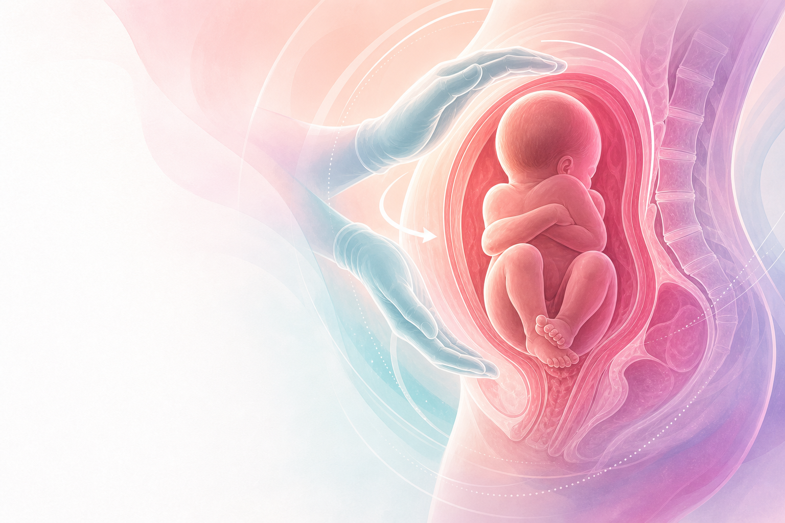
Clinical overview
Breech presentation — the fetus lying longitudinally with the buttocks or feet as the presenting part — is the commonest malpresentation at term, complicating roughly 3–4% of singleton pregnancies at 37 weeks (standard teaching). It is far commoner earlier in gestation (around a quarter of pregnancies before 28 weeks) because the relatively large fetal head naturally seeks the roomier uterine fundus until the fetus grows and spontaneously turns cephalic. Most breeches therefore correct themselves; the clinical problem is the minority that persist to term, and the decision about how that baby should be born.
The registrar must hold two parallel tasks in mind. Antenatally, the aim is to detect breech reliably (clinically and by ultrasound), exclude or define the reasons it is breech (placenta praevia, fibroids, fetal anomaly, oligohydramnios, multiple pregnancy), and offer the interventions that change the outcome — chiefly external cephalic version (ECV), which converts a breech into a cephalic presentation and so removes the risk altogether. Intrapartum, the task is to counsel honestly about mode of delivery, recognise that the evidence (the Term Breech Trial) shifted practice strongly towards planned caesarean, and yet retain the skills and judgement to conduct a safe vaginal breech birth when one is chosen or forced — the unbooked woman in advanced labour, the second twin, or the woman who declines surgery. In the South African context, where many women present late, undiagnosed breech in labour is common, and the ability to deliver a breech safely is not optional. This chapter sits alongside Complicated labour, Instrumental delivery and Multiple pregnancy.
Core knowledge
Types of breech
- Frank (extended) breech — hips flexed, knees extended, legs splinted up against the trunk. The commonest type at term (classically ~60–70%). The presenting part is well-applied to the cervix and the buttocks dilate the cervix effectively — the most favourable type for vaginal birth.
- Complete (flexed) breech — hips and knees both flexed; the fetus is "sitting cross-legged". Presenting part is buttocks and feet together.
- Footling (incomplete) breech — one or both hips extended so a foot is the presenting part. Carries the highest risk of cord prolapse (a small irregular presenting part poorly applied to the cervix) and is generally regarded as a contraindication to vaginal breech birth.
Why a breech is dangerous to deliver vaginally
The hazard of vaginal breech birth is mechanical and is concentrated in the after-coming head. In a cephalic birth the head — the largest, least compressible part — moulds and descends first, so by the time the body is born the pelvis has proven adequate. In a breech the soft, compressible buttocks and trunk deliver first through a cervix and pelvis that have not been tested by the head. The head then arrives last and rapidly, with no time to mould, and may become entrapped at an incompletely dilated cervix (especially if the baby is preterm, where the head is disproportionately large) or behind the symphysis. Once the cord is in the vagina alongside the trunk it is compressed, so there is a hard time limit on delivering the head before hypoxic injury. Other mechanisms of harm include cord prolapse (especially footling), nuchal arms (arms extended above the head, obstructing delivery), head extension/deflexion, and birth trauma to the brachial plexus, spine, abdominal viscera and genitalia from incorrect handling.
Figure J6.1 — Breech type and after-coming-head mechanics linking frank, complete and footling presentations to cord prolapse, head entrapment, nuchal arms, head extension and vaginal-birth selection criteria.
Causes and associations
A persistent breech is sometimes simply chance, but it should prompt a search for a reason that may itself change management:
- Reduced/abnormal fetal mobility or room to turn — oligohydramnios, fetal growth restriction (see Intrauterine growth restriction), fetal neuromuscular anomaly.
- Polyhydramnios — excessive mobility, late or unstable lie.
- Uterine/pelvic factors — uterine anomaly (septate, bicornuate), fibroids, previous breech.
- Placental — placenta praevia (a cause of malpresentation and an absolute contraindication to ECV and to vaginal birth — see Antepartum haemorrhage) or cornual placenta.
- Multiple pregnancy — restricted space; the breech second twin is a distinct scenario.
- Fetal — anomalies (notably hydrocephalus, anencephaly), and prematurity (the single commonest "association", since most preterm fetuses are breech).
Assessment
History and examination
The diagnosis is often first suspected on abdominal palpation at the antenatal visit: a firm, ballottable, round mass (the head) felt in the fundus; a softer, less defined presenting part in the pelvis; and the fetal heart auscultated higher than expected (at or above the umbilicus). Leopold's manoeuvres should be performed at every late antenatal contact; relying on the woman to report "feeling kicks low down" is unreliable. A high presenting part, an apparently small or oddly shaped abdomen, or difficulty identifying the lie should all trigger imaging.