Clinical overview
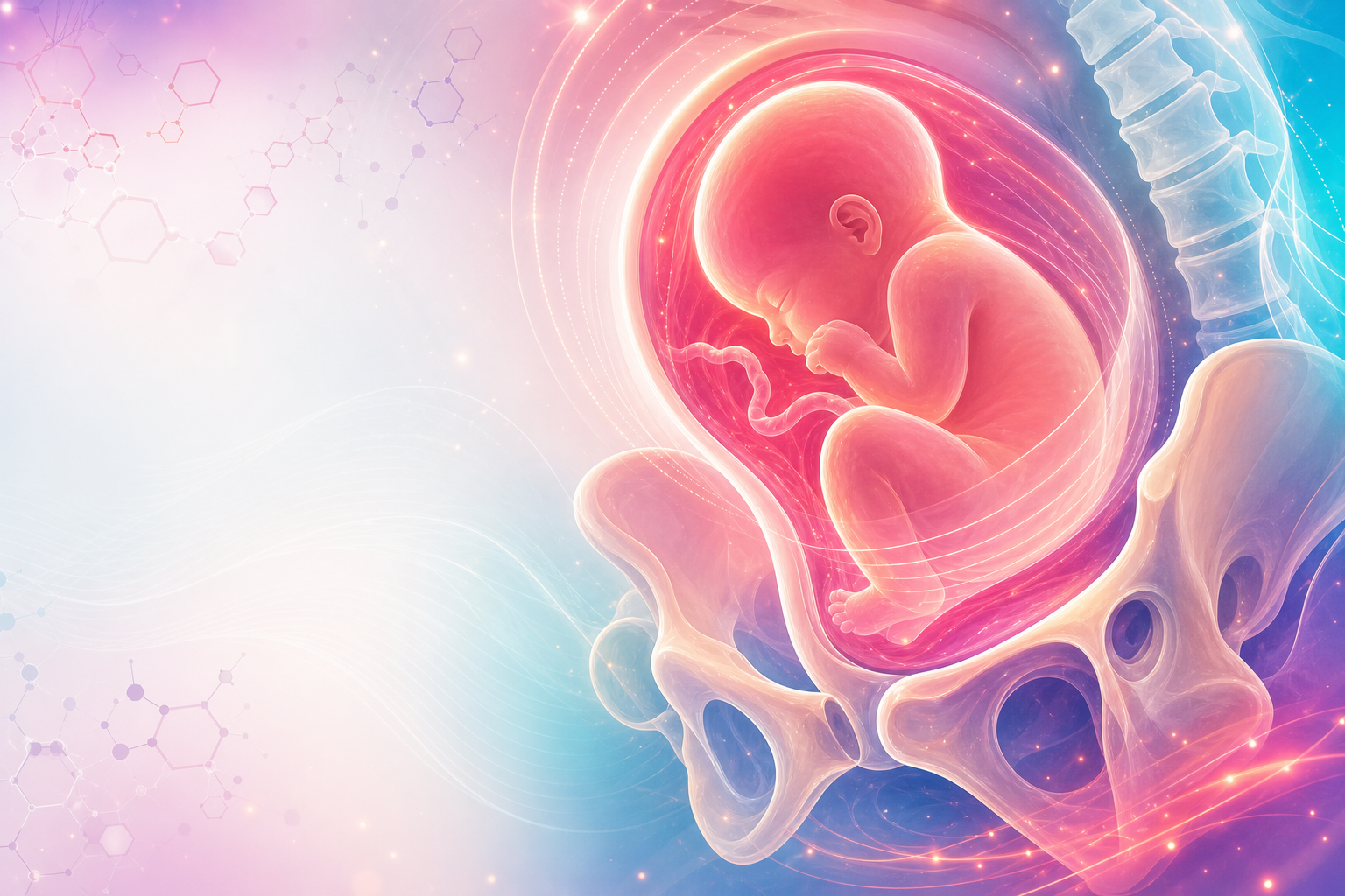
The macrosomic fetus is the large baby whose size threatens both itself and its mother — chiefly through obstructed and complicated labour, shoulder dystocia, birth trauma, perinatal asphyxia, and, on the maternal side, postpartum haemorrhage and severe perineal injury. The clinical problem is not the size itself but the mismatch between fetal dimensions and the maternal pelvis and soft tissues, played out under the time pressure of labour. Macrosomia therefore sits at the intersection of antenatal surveillance, intrapartum decision-making, and emergency obstetric drill.
For the FCOG(SA) registrar the central tension is one of prediction versus prevention. We cannot measure fetal weight precisely before birth — clinical estimation and ultrasound both carry wide error margins — yet we are asked to make high-stakes decisions (induction, elective caesarean, mode and place of delivery) on those imperfect estimates. Over-call it and you generate unnecessary caesareans, inductions, and maternal morbidity in a system where theatre access and blood are finite. Under-call it and you risk a shoulder dystocia in a district facility without the team or skills to manage it. In the South African setting — where undiagnosed and poorly controlled diabetes is common, where many women book late, and where the gradient between district, regional and tertiary care is steep — the macrosomic fetus is a recurring and high-consequence problem. This chapter frames how to suspect it, how to assess it honestly, how to plan delivery, and how to drill the emergency it most threatens: shoulder dystocia and PPH.
Core knowledge
Definitions
There is no single universal threshold, and the registrar must be able to state the candidate definitions and their rationale.
- Macrosomia classically refers to an absolute birth weight above a fixed cut-off, most commonly ≥4000 g, with ≥4500 g marking a higher-risk category, irrespective of gestational age. Some authorities add a ≥5000 g tier where morbidity rises steeply.
- Large for gestational age (LGA) is a relative definition: birth weight above the 90th centile for gestational age (and ideally for sex and population). A preterm infant can be LGA without being macrosomic by absolute weight.
These are not interchangeable. Absolute thresholds matter because the risks of shoulder dystocia and birth trauma climb with absolute fetal dimensions; centile-based definitions matter for identifying the pathological growth trajectory (especially the diabetic fetus) that warrants surveillance. Note the contrast with the small fetus covered in intrauterine-growth-restriction: both are growth-disorders, but macrosomia is over-growth with mechanical and metabolic consequences.
Pattern of growth — why the diabetic baby is different
Not all large babies are large in the same way, and this is examinable.
- Constitutionally large / symmetrical macrosomia: proportionate enlargement, often in tall parents or with grand multiparity and post-term pregnancy. Risk is mechanical (dystocia, trauma) but proportionate.
- Diabetic / asymmetrical macrosomia: driven by fetal hyperinsulinaemia. Maternal hyperglycaemia crosses the placenta; the fetal pancreas responds with insulin, which is a potent fetal growth factor acting on insulin-sensitive tissues. The result is disproportionate deposition of fat and glycogen in the trunk, shoulders and interscapular region, raising the shoulder-to-head and chest-to-head ratios. This is precisely the geometry that causes shoulder dystocia, which is why the diabetic macrosomic fetus carries a disproportionately higher dystocia risk at any given weight than the non-diabetic large baby.
Figure J22.1 — Macrosomia risk map showing absolute and centile definitions, diabetic asymmetrical growth, EFW uncertainty, and maternal-neonatal consequences.
Risk factors
- Maternal diabetes — pre-existing type 1/2 and gestational diabetes (GDM); the strongest modifiable driver, mediated by glycaemic control.
- Maternal obesity and excessive gestational weight gain.
- Previous macrosomic infant — strongly predictive of recurrence.
- Post-term pregnancy (continued growth beyond term).
- Multiparity and advanced maternal age; tall, heavy parents (genetic potential); male fetus.
- Rarely, fetal overgrowth syndromes (e.g. Beckwith–Wiedemann) — consider where macrosomia is extreme or syndromic features coexist.
Consequences
Fetal/neonatal: shoulder dystocia and its sequelae — brachial plexus injury (Erb's palsy), clavicular/humeral fracture, hypoxic-ischaemic injury and, in the worst case, intrapartum death; neonatal hypoglycaemia (hyperinsulinaemic, in the diabetic baby), polycythaemia, jaundice, and later-life metabolic risk.
Maternal: prolonged/obstructed labour, higher operative and caesarean delivery rates, severe perineal trauma including oasis, and postpartum-haemorrhage from an over-distended atonic uterus. The risk of uterine-rupture is relevant where macrosomia coexists with a scarred uterus or injudicious augmentation.
Assessment
History
Target the risk factors and prior obstetric record. Ask specifically about: previous large babies and their birth weights and any difficult delivery, shoulder dystocia, or neonatal injury; known or suspected diabetes and glycaemic control; booking weight and weight gain; symphysio-fundal height trends; and dates certainty (an over-estimate of gestation inflates apparent size). A documented previous shoulder dystocia is one of the most important single items in the chart — it materially raises recurrence risk and reframes the delivery plan.
Examination
- Symphysio-fundal height (SFH) plotted serially on the growth chart used in the antenatal record: an SFH measuring large-for-dates (classically >2–3 cm above gestational age in weeks, or crossing centiles) prompts further assessment but is a blunt screen, confounded by polyhydramnios, maternal habitus, multiple pregnancy and a full bladder.
- Clinical estimation of fetal weight (Leopold's manoeuvres / abdominal palpation) — useful, free, and the only tool available at many primary sites, but with a wide error margin, especially in the obese abdomen. Parous women's own estimate of "bigger than my last" deserves respect.
- Assess liquor volume clinically (polyhydramnios often accompanies the diabetic macrosomic fetus) and exclude multiple pregnancy as the cause of large-for-dates.
Investigations
- Ultrasound biometry to estimate fetal weight (EFW) and, importantly, to assess proportion (abdominal circumference relative to head circumference) and liquor volume. The registrar must internalise the central caveat: ultrasound EFW is imprecise at the upper extreme of weight, with typical errors of the order of ±10–15%, and it does not reliably predict shoulder dystocia. A large AC and polyhydramnios in a diabetic pregnancy are nonetheless meaningful flags. Never let a single confident-looking EFW number override clinical judgement — and never act on a number you would not be willing to defend as ±15%.
- Screen for and quantify diabetes in any large-for-dates pregnancy not already tested: glucose testing per the South African National Integrated Maternal and Perinatal Care Guideline (NDoH, 2024) pathway. Macrosomia or polyhydramnios discovered late should trigger glucose assessment even if earlier screening was normal or omitted (late booking is common). See medical-complications-in-pregnancy for diabetes in pregnancy in detail.
- Confirm gestational age against the earliest reliable dating (see gestational-age-assessment) — apparent macrosomia is sometimes simply mis-dating.
Management
Management spans the antenatal, intrapartum and postpartum periods, and the SA registrar must always layer the level-of-care and referral dimension onto the clinical plan.
Antenatal: prevention and planning
- Optimise glycaemic control — this is the single most effective lever to reduce excess fetal growth and is the reason diabetes detection and management dominate the antenatal plan. Tight, guideline-based glucose targets, dietary intervention, and pharmacotherapy where needed reduce the trajectory towards macrosomia.
- Counsel honestly about the limits of weight prediction and the options. The decision around suspected macrosomia is preference-sensitive and must be shared.
- Risk-stratify the delivery plan by level of care. A woman with suspected significant macrosomia — and especially one with diabetes, a previous shoulder dystocia, or a scarred uterus — should be delivered where there is immediate access to senior obstetric and neonatal skill, theatre and blood. In the SA tiered system this means planning delivery at an appropriately resourced regional/tertiary unit rather than a midwife-led district site, with timely antenatal referral so the plan is not made for the first time in established labour.
The mode-of-delivery decision
This is the HOTS heart of the objective. There is no single threshold that mandates caesarean, and the registrar must reason rather than recite a number.
- Elective caesarean section is reasonably offered and discussed at the highest weight estimates, where the absolute risk of catastrophic dystocia rises and the number-needed-to-section to prevent one permanent brachial plexus injury becomes less prohibitive. Standard teaching positions this discussion at EFW around ≥4500 g in the diabetic pregnancy and ≥5000 g in the non-diabetic pregnancy, but these are decision-aids for shared counselling, not automatic triggers — and they rest on an imprecise estimate. Caesarean for suspected macrosomia must be balanced against the maternal morbidity of operative delivery and the implications for future pregnancies in a setting where repeat caesarean and placenta accreta are real downstream costs.
- Induction of labour for suspected macrosomia at or near term, to deliver before further growth, is an option that may reduce shoulder dystocia and fracture risk in some settings, but the evidence is debated, it does not abolish dystocia, and it carries its own caesarean and intervention trade-offs. Decide case-by-case with the woman.
- Planned vaginal birth remains appropriate for many suspected-macrosomic pregnancies, particularly non-diabetic, with a favourable pelvis and no prior dystocia — but it must be vaginal birth with the team and skills ready for shoulder dystocia, conducted where escalation is immediate.
- A documented previous shoulder dystocia shifts the balance strongly: discuss elective caesarean explicitly, weighing severity of the prior event, EFW, glycaemic status and maternal preference.

Figure J22.2 — Delivery planning ladder for suspected macrosomia, linking assessment, level-of-care planning, counselling thresholds, induction, and vaginal-birth readiness.
Intrapartum care
- Deliver in a unit with senior obstetric presence, neonatal resuscitation capability and theatre/blood access. Anticipate, don't be surprised.
- Be cautious with augmentation. A labour that is slow with a clinically large baby may be telling you about disproportion; injudicious oxytocin in obstructed labour risks uterine-rupture, especially on a scar (see vbac).
- Maintain a low threshold for senior review and for abandoning a difficult instrumental attempt — mid-cavity instrumental delivery of a large baby that then arrests at the shoulders is a dangerous combination (see instrumental-delivery).
- In the diabetic labour, manage maternal glucose to reduce neonatal hypoglycaemia.
EMERGENCY DRILL — shoulder dystocia
Shoulder dystocia is an obstetric emergency. Recognise it, call for help, and work the sequence — do not pull harder.
The diagnosis is bony impaction of the anterior shoulder behind the symphysis after the head delivers: failure of restitution/external rotation, the "turtle-neck" sign (head retracts against the perineum), and failure of the shoulders to deliver with routine traction.
Run the drill, in order, without delay (the widely taught HELPERR structure):
- H — Call for HELP: declare "shoulder dystocia", summon the most senior obstetrician, additional midwives, an anaesthetist and the neonatal/resuscitation team; note the time.
- E — Evaluate for Episiotomy: consider one to make room for internal manoeuvres (the obstruction is bony, but it aids access).
- L — Legs (McRoberts' manoeuvre): hyperflex the maternal hips onto the abdomen — the single most effective first step, flattening the sacral promontory and rotating the symphysis.
- P — suprapubic Pressure: apply directed pressure behind the anterior shoulder (CPR-style) to adduct and disimpact it; combine with McRoberts'.
- E — Enter (internal rotational manoeuvres): rotate the shoulders to the oblique (e.g. Rubin II, Woods' screw / reverse Woods').
- R — Remove the posterior arm: deliver the posterior arm to reduce the diameter.
- R — Roll the patient onto all fours ("all-fours" / Gaskin manoeuvre) and repeat manoeuvres.
Avoid the three errors that injure babies and mothers: do not apply excessive downward axial traction (causes brachial plexus injury), do not apply fundal pressure (worsens impaction and risks rupture), and do not over-rotate the neck. Last-resort manoeuvres for the truly intractable case (cleidotomy, the Zavanelli cephalic replacement, symphysiotomy) are documented but are senior-only and rarely needed if the drill is performed promptly. Document contemporaneously: time of head and body delivery, sequence and timing of manoeuvres, which shoulder was anterior, traction used, cord gases, the personnel present and the neonatal condition — this record is clinically and medico-legally essential. SA units should drill shoulder dystocia regularly so the sequence is reflexive at 03:00 in a district labour ward.

Figure J22.3 — HELPERR shoulder dystocia drill with recognition signs, sequential manoeuvres, unsafe actions to avoid, and documentation points.
Postpartum
- Anticipate postpartum-haemorrhage: the over-distended uterus is prone to atony. Ensure active management of the third stage, oxytocin per protocol, IV access, and tranexamic acid early if PPH develops (TXA 1 g IV within 3 hours of onset). Have a haemorrhage plan ready.
- Inspect carefully for oasis and severe perineal/vaginal trauma and repair appropriately.
- Neonatal care: assess for birth injury (brachial plexus, fractures), and screen the diabetic-exposed baby for hypoglycaemia with early feeding and glucose monitoring; involve neonatology where needed. Provide neonatal-resuscitation readiness for any difficult birth.
- Debrief the mother and the team after a shoulder dystocia, and counsel on recurrence and the plan for any future pregnancy.
Red flags / pitfalls
- Trusting the ultrasound EFW as if it were exact. It is ±10–15% at the top end and does not predict dystocia. Acting on a single decimal-point number is a recurring error — never let it override clinical judgement or, conversely, falsely reassure.
- Forgetting that the diabetic large baby is more dangerous per gram — disproportionate shoulder/trunk fat means dystocia risk outstrips weight alone. A "borderline" EFW in a poorly controlled diabetic is not reassuring.
- Missing late or undiagnosed diabetes. New large-for-dates or polyhydramnios must trigger glucose testing even after a "normal" earlier screen or a late booking.
- Pulling harder in shoulder dystocia. Traction and fundal pressure cause the very injuries the drill exists to prevent. Work the sequence; never haul.
- Augmenting an obstructed labour. Slow progress with a clinically large baby may be disproportion; oxytocin then risks uterine-rupture, catastrophically on a scar.
- Planning delivery in the wrong place. A suspected macrosomic, diabetic, or previous-dystocia patient delivering at a district midwife-led site without theatre, blood or senior skill is a system failure — refer antenatally so the plan is in place before labour.
- Over-medicalising the constitutionally large baby. Not every big baby needs induction or caesarean; balance dystocia risk against the maternal morbidity of intervention, including future-pregnancy consequences of caesarean.
- Inadequate documentation of a shoulder dystocia — a frequent and serious deficiency with clinical and medico-legal consequences.
Evidence anchors
- South African National Integrated Maternal and Perinatal Care Guideline (NDoH, 2024) — the SA obstetric source of truth: antenatal care, diabetes screening pathway, levels of care and referral, intrapartum management.
- NICE NG3 — Diabetes in Pregnancy — glycaemic targets and management of the diabetic pregnancy that drive macrosomia prevention; informs mode-of-delivery counselling for the diabetic large baby.
- RCOG Green-top Guideline No. 42 — Shoulder Dystocia — recognition, the manoeuvre sequence, avoidance of traction/fundal pressure, and documentation standards underpinning the emergency drill above.
- NICE NG235 — Intrapartum Care and NICE NG229 — Fetal Monitoring in Labour — intrapartum monitoring and care of the complicated labour.
- Saving Mothers (NCCEMD) report — obstetric haemorrhage and the burden of complicated delivery in SA, supporting the PPH anticipation and referral emphasis.
- WOMAN trial (Lancet 2017) — tranexamic acid 1 g IV within 3 hours of PPH onset reduces death from bleeding; underpins early TXA in the over-distended, post-macrosomic uterus.
- ILCOR 2025 / NRP — neonatal resuscitation readiness for the difficult or injured birth.
Author's note on hedged facts: the weight thresholds for offering caesarean (≈≥4500 g diabetic / ≥5000 g non-diabetic), the ≥4000 g/≥4500 g macrosomia tiers, the ultrasound EFW error margin (±10–15%), and the SFH ">2–3 cm" rule are stated as standard teaching / decision-aids rather than as line-itemed thresholds in VERIFIED-SOURCES.md, and are written cautiously here. Specific glycaemic target numbers and the precise SA diabetes-screening algorithm should be confirmed against the NDoH 5th-edition (2024) and NICE NG3 source documents before being quoted as exact figures.
