Clinical overview
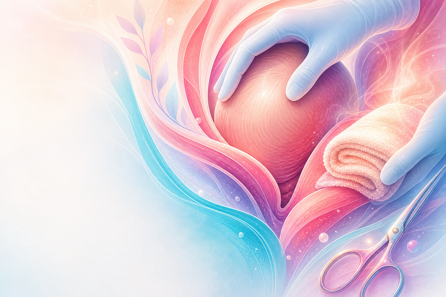
The perineum is the soft-tissue bridge between vagina and anus that every vaginal birth must negotiate. Most parturients sustain some degree of perineal trauma at first vaginal birth, and a clinically important minority sustain obstetric anal sphincter injury (OASIS) with lifelong consequences for continence, sexual function and psychological wellbeing. The registrar's task is rarely a single dramatic intervention; it is a sequence of small, evidence-graded choices across the second stage — how the head is allowed to deliver, whether and where a hand is placed, whether a warm compress is applied, whether and when an episiotomy is cut, and how completely any tear is then diagnosed and repaired. Each choice shifts the probability of an intact perineum or of a third- or fourth-degree tear by a few percentage points; together they are the difference between a woman who walks out continent and one referred to a colorectal clinic.
This objective asks you to appraise — to weigh competing techniques rather than simply recite a protocol. That weighing matters acutely in the South African setting, where most births occur in midwife-led labour wards at district level, where instrumental delivery and macrosomia are common, and where the recognition and primary repair of OASIS often falls to a registrar called from another ward. The National Integrated Maternal and Perinatal Care Guideline (NDoH, 2024) frames perineal care within respectful, woman-centred intrapartum care; getting it right is both a quality marker and a medicolegal flashpoint. This chapter sits alongside normal-labour, instrumental-delivery and oasis, and the techniques below assume you have already mastered the mechanics of the normal second stage.
Core knowledge
Anatomy of the perineum
The perineal body is a fibromuscular pyramid into which the bulbospongiosus, superficial and deep transverse perineal muscles, the external anal sphincter (EAS) and fibres of the levator ani converge. Understand the layered structure because repair recapitulates it. From the introitus outward and downward: vaginal epithelium and submucosa; the perineal muscles and the perineal body; subcutaneous tissue and perineal skin. Posterior to the perineal body lie the EAS (a circular striated voluntary muscle), the internal anal sphincter (IAS, a smooth-muscle continuation of the rectal circular muscle — pale, glistening, and the key to continence of flatus and liquid stool) and the anorectal mucosa. See genital-anatomy for the full pelvic floor.
Classification of perineal trauma
The internationally accepted (RCOG/Sultan) classification grades severity by depth:
| Degree | Structures involved |
|---|---|
| First | Perineal/vaginal skin and mucosa only |
| Second | Perineal muscles, sphincter intact (includes most episiotomies) |
| Third (3a) | EAS torn, <50% of its thickness |
| Third (3b) | EAS torn, >50% of its thickness |
| Third (3c) | Both EAS and IAS torn |
| Fourth | EAS, IAS and anorectal mucosa torn |
Third- and fourth-degree tears together constitute OASIS. A "buttonhole" tear (rectal mucosa torn with an intact sphincter) is a separate entity and is easily missed. Anterior trauma to labia, urethra and clitoris is graded separately and often left unsutured if not bleeding and well-apposed. The classification and the principles of OASIS repair are anchored in RCOG Green-top Guideline No. 29; this chapter deliberately defers the detail of OASIS recognition and repair to oasis.
Risk factors for severe trauma
Recognising the high-risk birth lets you mobilise prevention. Classically described associations include nulliparity, larger fetal weight/macrosomia (see macrosomia), instrumental delivery (especially forceps), occipito-posterior position, prolonged second stage, shoulder dystocia, and previous OASIS. Asian ethnicity is a frequently cited risk factor in the OASIS literature. Many of these are non-modifiable on the day; what you can modify is the conduct of the delivery itself.
Assessment
Antenatal and intrapartum appraisal
Identify the woman at elevated risk during labour: the primigravida with an estimated large baby, the persistent OP position, the instrumental birth in prospect, and crucially the woman with a previous OASIS — whose mode of delivery and perineal plan should already have been discussed and documented antenatally, with counselling about recurrence risk and the option of elective caesarean for the subtotal-incontinent or symptomatic woman.
Examining the perineum after birth
Systematic post-delivery examination is the single most important step in not missing OASIS, and it is where registrars are most often found wanting in medicolegal review. Every woman after vaginal birth — including those who appear to have an intact perineum — must have a structured genital and rectal examination. Standard teaching, reflected in RCOG GTG 29, is:
- Adequate analgesia and lighting, with the woman in a position allowing a clear view (lithotomy where available).
- Inspect the full extent of the tear — apex of any vaginal laceration, the perineal muscles, and the anal margin.
- Perform a digital rectal examination to feel for a sphincter defect (the "torn end retracts laterally") and to exclude a buttonhole injury, before deciding the tear is second-degree.
- Use the "pill-rolling" palpation of the EAS and ask the woman to squeeze if she is awake and cooperative.
- Classify explicitly and document the degree, the structures involved, and who performed the exam.
If there is any doubt about sphincter integrity, treat as OASIS and escalate — under-classification (calling a 3a a "second-degree") is the commonest and most consequential error. Examination under regional or general anaesthesia in theatre is appropriate where exposure or analgesia at the bedside is inadequate.
Diagnosing and quantifying blood loss
The perineum and lower vagina bleed briskly; perineal and vaginal trauma is a recognised contributor to primary postpartum-haemorrhage. Inspect the apex of every vaginal tear (bleeding can track upward and be concealed) and quantify loss as part of the standard PPH response.
Management
This is the heart of the objective: appraising the means of protecting the perineum, then the use of episiotomy, then repair of tears.
Protecting the perineum during the second stage
No single manoeuvre abolishes trauma; the evidence supports a bundle of behaviours that together reduce OASIS rates. The components with the best support are:
1. Controlled, slow delivery of the head. Allowing the head to deliver slowly between contractions — guarding against an explosive crowning — gives the perineum time to stretch and is the conceptual core of most protective techniques. Communicate with the woman: ask her to stop active pushing and to pant or "breathe the baby out" as the head crowns.
2. Manual perineal protection ("hands-on"). The classic technique places one hand on the fetal occiput to control the speed of the head while the other (thumb and fingers) supports the perineum, sometimes flexing the head. The competing "hands-poised" (or hands-off) approach keeps the hands ready but does not touch. The evidence here is genuinely contested: large trials and meta-analyses have not shown a consistent, decisive advantage of one over the other for OASIS, and standard teaching is that either is acceptable provided the head is delivered slowly and under control. The pragmatic registrar position — reflected in OASIS-prevention bundles such as the UK/RCOG-associated care bundle — is to use a controlled hands-on technique by default while supporting maternal-led pushing, and not to dogmatically insist on either label. (The relative efficacy of hands-on vs hands-poised is not a settled, line-itemed verified guideline statement — present it as contested.)
3. Warm perineal compress. A warm compress applied to the perineum during the late second stage is one of the better-supported single interventions for reducing third- and fourth-degree tears and is cheap, acceptable and feasible in a district labour ward. (Quantified effect sizes vary across trials — standard teaching is a reduction in severe tears; treat the magnitude cautiously.)
4. Position and pushing. There is no strong evidence mandating a particular maternal position to prevent OASIS; flexibility and maternal preference are appropriate, consistent with the respectful-care ethos of the NDoH 5th edition guideline and respectful-care. Avoid sustained directed (Valsalva) pushing where spontaneous pushing is effective.
5. Perineal massage — antenatal perineal self-massage from ~34–35 weeks is associated with a modest reduction in trauma requiring suturing in primiparous women and can be offered. Intrapartum massage during the second stage has weaker and more variable support.
The pragmatic appraisal for the exam: warm compress, slow controlled delivery of the head and good communication are the evidence-favoured, low-cost interventions to do; hands-on vs hands-off is a technique choice, not a quality marker, provided control of the head is maintained.
Figure J9.1 — Perineal protection bundle for the second stage: risk scan, warm compress, controlled crowning, flexible hands-on or hands-poised support, and post-birth examination before classifying trauma.
Use of episiotomy
Episiotomy is a deliberate incision of the perineum to enlarge the outlet. Its appraisal is one of the cleanest examples in obstetrics of how a once-routine intervention became selective.
Restrictive, not routine. The pivotal lesson — now embedded in WHO and NICE intrapartum guidance (and SA practice) — is that routine episiotomy does not protect the perineum and increases the rate of posterior trauma; episiotomy should be used selectively/restrictively, for a clinical indication, not for every primigravida. Recognised indications include suspected fetal compromise requiring expedited delivery, instrumental birth (particularly forceps), and an anticipated severe tear or shoulder dystocia where outlet room is needed.
Mediolateral, not midline. Where an episiotomy is indicated, the mediolateral incision is preferred in modern UK/SA practice because the midline incision, though it heals well and is less painful, carries a substantially higher risk of extension into the anal sphincter (OASIS). The recommended technique:
- Adequate analgesia first — infiltrate with local anaesthetic (e.g. lidocaine) if no effective regional block is in place; do not cut an un-anaesthetised perineum.
- Cut at crowning, when the perineum is maximally stretched and thinned, distending over the fetal head.
- Direct the incision from the posterior fourchette at an angle of roughly 60° from the midline (so that the angle is closer to 45° once the perineum retracts post-delivery), aiming away from the anal sphincter. A too-vertical (near-midline) mediolateral cut loses its protective rationale.
- Use sharp scissors in a single deliberate cut at the height of a contraction.
(The 60°-at-crowning angle and the midline-vs-mediolateral OASIS risk are standard teaching consistent with RCOG GTG 29 / NICE intrapartum guidance; the precise degree figure should be taught as "approximately 60°" rather than as a hard verified threshold.)

Figure J9.2 — Episiotomy decision board contrasting restrictive clinical indications with routine cutting, and showing the mediolateral angle, timing and analgesia checks that reduce sphincter-extension risk.
Repair of perineal tears
First-degree and superficial trauma. Skin-only first-degree tears that are well-apposed and not bleeding may be left unsutured; suture if bleeding or malaligned. Use a fine absorbable suture.
Second-degree tears and episiotomy are repaired in layers under adequate analgesia, with the same principles:
- Identify the apex of the vaginal tear first and place the first suture above it to secure a retracted bleeding vessel.
- Repair in three layers: a continuous non-locking suture to the vaginal wall down to the fourchette; interrupted or continuous sutures to approximate the perineal muscles (deep then superficial); then a continuous subcuticular suture to the perineal skin (associated with less short-term pain than interrupted transcutaneous sutures).
- Use a rapidly-absorbed polyglactin (e.g. Vicryl Rapide) type suture for the perineal skin/muscle — standard teaching and consistent with NICE intrapartum recommendations — to reduce suture-removal need and dyspareunia.
- Count swabs and needles before and after; perform a rectal examination at the end of every repair to confirm no suture has been passed through the rectal mucosa.
OASIS (third- and fourth-degree) must be repaired by an appropriately trained clinician in theatre under regional/general anaesthesia with good lighting and assistance — not on the labour-ward bed. The detail (overlap vs end-to-end EAS repair, IAS repair, antibiotics, laxatives, follow-up) is covered in oasis. The non-negotiable principle for this chapter: recognise it, do not under-classify it, and escalate it to theatre.
South African context
- Level of care. Most vaginal births occur at district (Level 1) facilities, frequently midwife-led. Uncomplicated first- and second-degree repair is within midwifery and medical-officer scope; OASIS repair requires escalation — to a doctor on site able to repair in theatre, or referral/transfer to a regional (Level 2) hospital where bedside expertise and theatre access are lacking. Know your facility's pathway before you need it.
- NDoH 5th-edition guideline frames perineal care within respectful intrapartum care: informed, consented examination (including the rectal exam), adequate analgesia for repair, and clear documentation.
- Resource realities. Suture material, lighting and theatre access vary; a warm compress and slow controlled delivery are interventions available everywhere and should be standard. Where lithotomy and good lighting are not available at the bedside, examine and repair in theatre rather than risk missing OASIS.
- HIV. A high proportion of parturients are HIV-positive; the relevant intrapartum concern is staff sharps safety (needlestick during repair) and the woman's ART/PMTCT status per hiv-in-pregnancy — there is no routine modification of perineal repair technique for maternal HIV status, but adopt universal precautions and a no-touch suturing technique.
- Audit. OASIS rate is a Saving Mothers/perinatal-care quality marker; under-detection (a falsely low rate) is as concerning as a high rate.
The emergency: when perineal trauma is haemorrhage
A briskly bleeding vaginal or perineal tear can be a life-threatening primary PPH while the uterus is well contracted. Make the drill unmistakable:
- Call for help and start the PPH drill — do not work alone on a bleeding tear in poor light.
- Apply direct pressure to the bleeding point and confirm the uterus is contracted (exclude atony as the dominant cause; give a uterotonic per protocol if any doubt).
- Get exposure: good lighting, lithotomy, an assistant, suction — move to theatre if the apex cannot be seen or the bleeding is not controlled at the bedside.
- Secure the apex first with a suture placed above it; a retracted arterial bleeder above an unidentified apex is the classic trap.
- Resuscitate in parallel — IV access, fluids, bloods/crossmatch, and tranexamic acid 1 g IV as part of the PPH bundle (give early, within 3 h of onset). (TXA dose/timing per the WOMAN trial and SA PPH guidance.)
- Re-examine and document, and exclude a concealed upper-vaginal or cervical tear if bleeding persists despite a repaired perineum.

Figure J9.3 — Post-birth perineal trauma drill linking structured genital and rectal examination, tear-depth classification, repair escalation, and the primary PPH response to a briskly bleeding tear.
Red flags / pitfalls
- Under-classifying OASIS. Calling a third-degree tear "second-degree" because no rectal exam was done is the single most damaging and most litigated error. Do a rectal exam on every vaginal birth.
- Missing the apex. Failing to identify and suture above the apex of a vaginal tear → ongoing concealed bleeding and PPH.
- Missing a buttonhole injury — rectal mucosa torn with an intact sphincter; suspect it, look for it.
- Routine episiotomy — cutting every primigravida is outdated and harmful; episiotomy is selective.
- Midline mediolateral confusion — a "mediolateral" cut directed too close to the midline loses its sphincter-sparing rationale; aim ~60° at crowning.
- Cutting too early or too late — an episiotomy cut before crowning bleeds more and is poorly placed; cut at crowning over the distending head.
- Inadequate analgesia for examination or repair — never cut or suture an un-anaesthetised perineum.
- Repairing OASIS on the labour bed in poor light by an untrained operator — escalate to theatre and a trained clinician.
- Forgetting the swab/needle count and final rectal exam — a suture through the rectal mucosa causes a fistula.
- Treating perineal bleeding as "just a tear" when it is a primary PPH — escalate early.
Evidence anchors
- National Integrated Maternal and Perinatal Care Guideline, South Africa (NDoH, 2024), NDoH — the SA obstetric source of truth; respectful intrapartum care, perineal examination/repair, levels of care and referral.
- RCOG Green-top Guideline No. 29 — Third- and Fourth-degree Perineal Tears (OASIS) — classification, structured examination including rectal exam, and repair principles (detail in oasis).
- RCOG Green-top Guideline No. 26 — Assisted Vaginal Birth — episiotomy in the context of instrumental delivery.
- NICE NG235 — Intrapartum care (2023) — restrictive/selective episiotomy, mediolateral technique, perineal repair (apex first, continuous subcuticular skin closure, rapidly-absorbed polyglactin), and post-repair rectal examination.
- WHO intrapartum recommendations / WHO Labour Care Guide (2020) — selective rather than routine episiotomy; woman-centred, respectful second-stage care.
- RCOG GTG 52 — Prevention and Management of Postpartum Haemorrhage and the WOMAN trial (Lancet 2017) — tranexamic acid 1 g IV early for trauma-related PPH.
- Saving Mothers / NCCEMD reports — obstetric haemorrhage as a leading SA maternal-death cause and the relevance of perineal/genital-tract trauma; OASIS detection as a perinatal-care quality marker.
