In one line
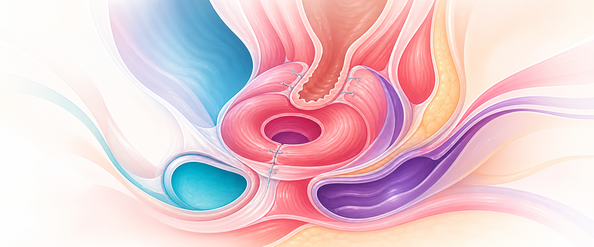
Obstetric anal sphincter injury is a continence disaster manufactured in the delivery room, so the controllable outcome lives almost entirely in two places — preventing the tear and recognising it the moment it happens — because a sphincter repaired correctly the first time does far better than any salvage, and most post-obstetric faecal incontinence is a missed or mis-repaired OASIS presenting late.
Mechanism & pathophysiology
Continence is a layered system, and faecal incontinence after childbirth almost always means one or more of those layers has failed in a way that maps directly onto a treatment. Four components hold flatus, liquid and solid stool. The internal anal sphincter (IAS) — smooth, involuntary, the terminal continuation of the rectal circular muscle — generates roughly 70–85% of resting anal tone and is the structure that quietly defends against passive soiling and flatal leakage. The external anal sphincter (EAS) — striated, voluntary, tonically active, pudendal-innervated — supplies the squeeze pressure that defers defaecation when the rectum fills. The puborectalis sling pulls the anorectal junction forward into the anorectal angle, a mechanical flap-valve that resists the rise in intra-abdominal pressure. Rectal compliance and sensation allow the rectum to accommodate a bolus and to discriminate flatus from stool. Strip any one component and a different incontinence phenotype appears: lose the IAS and you get passive soiling; lose the EAS and you lose deferment (urge incontinence of stool); blunt rectal sensation and the woman is soiled before she knows the rectum is full.
The groundwork for the injury itself — sphincter anatomy, the Sultan classification, the mechanics of the tear at crowning, the risk-factor list — is assumed here; revise it at the Intermediate OASIS chapter. What the consultant must hold is why obstetric faecal incontinence has two mechanistically distinct sources, because they respond to different operations.
Structural injury is a mechanical defect in the muscle ring — the torn sphincter of an acute OASIS, or the residual defect of one that healed badly. This is the lesion that endoanal ultrasound shows and that a sphincteroplasty can, in principle, close. Neuropathic injury is denervation of an anatomically intact sphincter, classically a pudendal neuropathy from stretch and compression during a prolonged, obstructed or instrumental second stage (and compounded over a lifetime by repeated vaginal delivery and chronic straining). The two coexist far more often than either occurs alone, which is the single most important pathophysiological fact for prognosis: a woman with a "successful" anatomical sphincter repair who still leaks usually has an unrecognised neuropathic component that no further surgery will fix. It also explains the cruel natural history of secondary sphincteroplasty — early results are good, then continence decays over five to ten years as an ageing, partly denervated muscle fatigues.
Occult OASIS is the third mechanistic strand and the reason endoanal ultrasound matters. A proportion of women who are recorded as having an intact perineum or a second-degree tear in fact have a sphincter defect demonstrable on imaging — an injury that was real at delivery but never seen, either because the rectal examination was never done or because the defect was sub-clinical at the time. These occult defects are a major contributor to the prevalence of faecal incontinence in parous women presenting years later, and they reframe the delivery-room examination from a formality into the primary preventive act of the whole topic.
A second mechanism deserves explicit attention because it changes the operation: the posterior compartment does not begin and end at the sphincter. The same delivery forces that tear the sphincter also stretch and disrupt the rectovaginal septum and perineal body, and a deficient perineal body — a foreshortened, attenuated central tendon — leaves the anorectum poorly supported even when the sphincter ring is intact. Clinically this presents as a combination of obstructed defaecation, a sensation of incomplete emptying, a posterior vaginal bulge (rectocele) and, on a background of sphincter weakness, frank incontinence. The consultant point is that "faecal incontinence after childbirth" is rarely a pure sphincter problem; it is frequently a compartment problem in which sphincter injury, perineal-body loss and rectal-wall prolapse coexist, and a repair that addresses only the sphincter while ignoring a deficient perineal body or an obstructing rectocele will under-deliver. This is why the posterior-compartment assessment — perineal-body length, evidence of rectocele or rectal intussusception, and defaecatory symptoms — belongs inside the faecal-incontinence work-up rather than alongside it.
The mechanistic synthesis that should drive every later decision is therefore a triage by which layer has failed: a structural EAS/IAS defect (sphincteroplasty-amenable, ultrasound-visible), a neuropathic but anatomically intact sphincter (better served by neuromodulation than by the knife), a deficient perineal body or rectocele (a reconstructive rather than a sphincter problem), and the very common situation in which several of these overlap. Reasoning back from the failed layer to the operation is what separates a durable result from a disappointing one.
Assessment
The assessment splits cleanly into two settings — the woman on the delivery bed minutes after birth, and the woman in clinic months or years later — and the consultant errors differ in each.
At delivery, every vaginal birth ends with a systematic perineal and digital rectal examination, not only the ones that look traumatic. The injury is missed when the examiner stops at the visible vaginal tear. With adequate analgesia and lighting:
- Inspect the whole perineum, vagina and the apex of any tear or episiotomy.
- Perform a per-rectum examination — a finger in the anal canal, a thumb in the vagina (or direct visualisation), palpating the sphincter while the woman squeezes if the block allows. Feel for the gap or "torn drawstring" of the EAS and the paler, firmer IAS beneath the pink mucosa.
- Grade precisely by the Sultan/RCOG system — 3a (<50% EAS thickness), 3b (>50% EAS), 3c (EAS and IAS torn), fourth-degree (EAS + IAS + anorectal mucosa) — because the grade drives the repair and the counselling.
- Look separately for a buttonhole rectal tear with an apparently intact sphincter, which predisposes to fistula and is repaired in its own right.
The commonest and most expensive assessment failures are not exotic: a 3a called a second-degree tear, and an unrecognised IAS injury inside a tear that was identified as third-degree. Both are downgrades of a real injury, and both surface later as incontinence.
In the symptomatic woman later, the assessment is the colorectal–urogynaecology one:
- A structured continence history that quantifies the problem and separates the phenotypes — passive soiling versus urge incontinence, flatus versus liquid versus solid, pad use, nocturnal leakage, the effect on work and sexual function. Use a validated severity score — the St Mark's (Vaizey) or Cleveland Clinic (Wexner) score — both to grade severity and to give an objective baseline against which any intervention is measured.
- Endoanal ultrasound maps the sphincter anatomy: it identifies or excludes a repairable structural defect and is the investigation to do when sphincteroplasty is being considered. A normal ring in a woman with intractable incontinence argues against a structural cause rather than diagnosing denervation, and she will not be helped by a sphincteroplasty.
- Anorectal manometry quantifies resting pressure (IAS function) and squeeze pressure (EAS function). Pudendal nerve terminal motor latency is not routinely recommended, because it does not reliably predict the outcome of a sphincter repair or of neuromodulation (ASCRS 2023); neurophysiology has only a limited, case-specific role. These are specialist, tertiary-level investigations in the SA context; they are not needed to recognise an acute OASIS, which stands entirely on the clinical examination.
- Defaecating proctography or dynamic MRI is reserved for the woman whose symptoms point to the posterior compartment — obstructed defaecation, incomplete emptying, a vaginal bulge — to demonstrate a rectocele, rectal intussusception or perineal descent, because these change the operation from a sphincter repair to a reconstruction. They are not part of the routine incontinence work-up and are tertiary investigations.