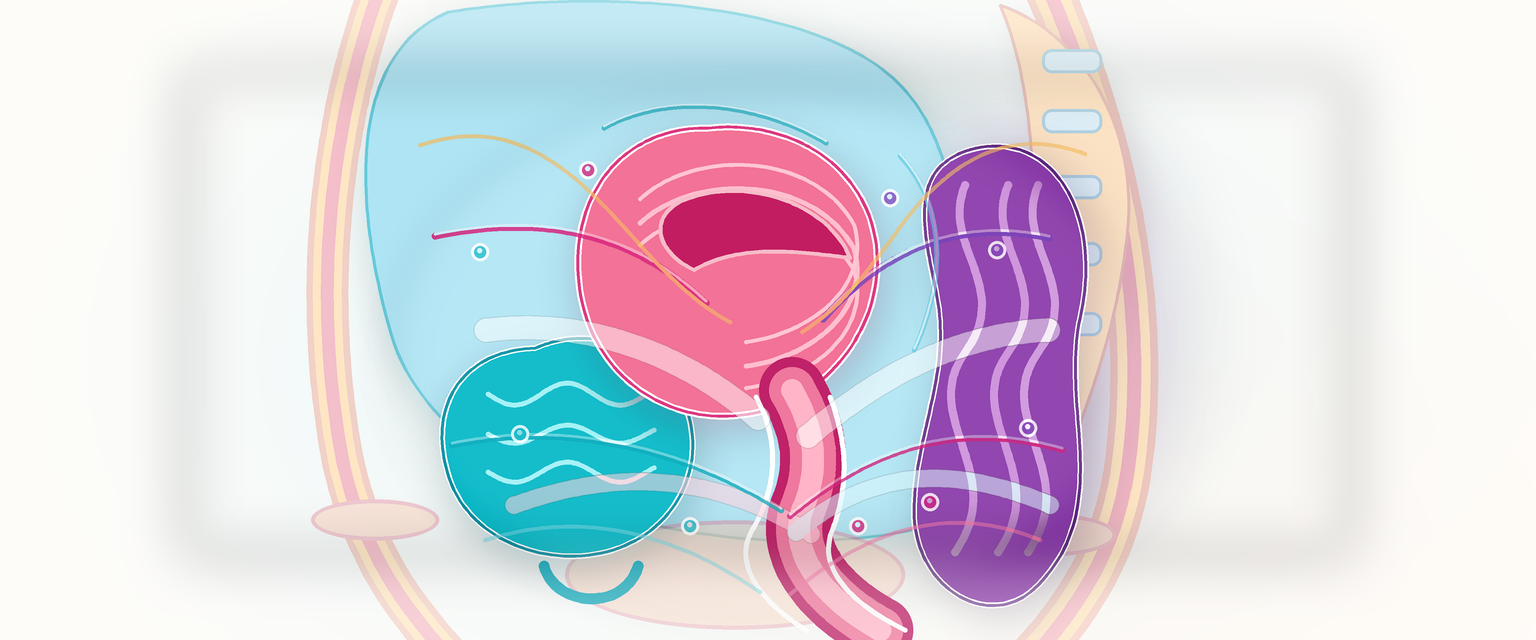
Pelvis: Bones, Viscera, Supports, Nerves, Vessels and Lymphatics
Pelvic anatomy is the operating map of obstetrics and gynaecology. The bony pelvis determines the passage through which the fetal head must descend and rotate. The pelvic floor and endopelvic connective tissue determine continence and prolapse risk. The ureter, uterine artery, internal iliac vessels, obturator nerve, pudendal nerve and autonomic plexuses determine whether surgery is safe. Lymphatic drainage determines cancer spread. Pain pathways determine why uterine contraction, cervical dilation, ovarian torsion and perineal stretching feel different.
The correct model is a layered cylinder:
- a bony ring with inlet, midpelvis and outlet;
- pelvic walls lined by obturator internus, piriformis and fascia;
- viscera arranged anterior to posterior: bladder/urethra, uterus/cervix/vagina, rectum/anal canal;
- vessels, ureter, nerves and lymphatics running in sidewall and parametrial tissue;
- a pelvic floor closing the outlet;
- the perineum below the pelvic floor.
Orientation: a Pelvic Cross-Section in Words
At the pelvic brim, the ureters cross the iliac vessels and enter the pelvis. On the sidewall, the external iliac vessels run toward the inguinal ligament; the internal iliac vessels dive into the pelvis; the obturator nerve runs along the lateral wall toward the obturator canal; the ovary lies in the ovarian fossa; the infundibulopelvic ligament carries ovarian vessels from the abdominal aorta. In the midline, the bladder sits anteriorly, uterus and cervix centrally, rectum posteriorly. The cervix projects into the vagina, creating fornices. The posterior fornix lies close to the pouch of Douglas.
| Structure | Where to picture it | Why it matters |
|---|---|---|
| Ureter | Retroperitoneal, sidewall, under uterine artery, into bladder | Hysterectomy, adnexal surgery, caesarean extension |
| Uterine artery | Base of broad ligament to cervix and uterus | Haemorrhage, hysterectomy pedicle, PPH ligation |
| Ovarian vessels | Infundibulopelvic ligament | Oophorectomy, torsion, para-aortic lymph drainage |
| Obturator nerve | Lateral wall, obturator fossa | Lymphadenectomy injury, medial-thigh pain |
| Pudendal nerve | Around ischial spine into Alcock canal | Pudendal block, perineal pain, sphincter function |
| Levator ani | Muscular pelvic floor | Prolapse, continence, childbirth injury |
| Cardinal/uterosacral complex | Parametrial/apical support | Apical prolapse, hysterectomy danger zone |
The pelvis is not a flat drawing. Most mistakes occur when a candidate memorises a named relation but cannot place it in depth: peritoneum, retroperitoneum, sidewall, parametrium, vaginal fornix and pelvic floor are different planes.
Bony Pelvis and Obstetric Mechanics
Start with the bones themselves. The bony pelvis is a ring formed by the two hip bones in front and at the sides, and the sacrum and coccyx behind. Each hip bone (os innominatum) is made from three bones — ilium, ischium and pubis — that are joined by cartilage in childhood and fuse in adulthood at a Y-shaped junction within the acetabulum. The sacrum is five fused vertebrae; it articulates with the fifth lumbar vertebra above, the coccyx below and the ilium on each side at the sacroiliac joints. Obstetrically, the pelvis is divided into the false pelvis above the brim and the true pelvis below it. The fetal head must pass through the true pelvis.
The pelvic ring is held together by three joints — the two sacroiliac joints posteriorly and the pubic symphysis anteriorly — and by strong ligaments. These joints move very little in the non-pregnant state, but in pregnancy the hormone relaxin and high progesterone soften the ligaments and cartilage, so the joints loosen to widen the available diameters during birth. The price of that adaptive laxity is pelvic girdle pain (formerly "symphysis pubis dysfunction"), which ranges from mild discomfort to disabling instability. The clinical relevance is concrete: when a woman has symptomatic pelvic girdle pain, forced or wide leg abduction — easy to do unintentionally once an epidural removes the pain warning — can worsen a symphyseal separation. Documenting a comfortable knee-to-knee abduction distance before labour protects the joint when she can no longer feel it.
The pelvic inlet is bounded by the sacral promontory, sacral ala, arcuate line, pectineal line, pubic crest and upper border of the pubic symphysis. It is usually widest transversely. This is why the fetal head commonly enters the brim in a transverse or oblique diameter.
The midpelvis is the level of the ischial spines. It is the level at which station is assessed, the level at which arrest often declares itself, and the landmark for pudendal block. The pelvic outlet is bounded by the lower pubic symphysis, pubic arch, ischial tuberosities, sacrotuberous ligaments and coccyx; it is widest anteroposteriorly.
| Level | Shape and landmarks | Obstetric meaning |
|---|---|---|
| Inlet | Transversely oval; sacral promontory to pubis | Engagement and entry diameter |
| Midpelvis | Around ischial spines; sidewalls and sacrum | Station, rotation, arrest, pudendal block landmark |
| Outlet | AP oval; pubic arch, tuberosities, coccyx | Crowning, outlet obstruction, perineal stretch |
Approximate obstetric dimensions are worth understanding rather than reciting:
| Pelvic level | Transverse | Oblique | Anteroposterior |
|---|---|---|---|
| Inlet | 13 cm | 11 cm | 11 cm |
| Midpelvis | 12 cm | 12 cm | 12 cm |
| Outlet | 10.5 cm | 11.5 cm | 12.5 cm |
The anteroposterior diameter of the inlet has named subtypes that an O&G specialist must keep straight, because they are how the inlet has historically been assessed clinically:
- The true (obstetric) conjugate runs from the sacral promontory to the nearest point on the upper border of the pubic symphysis. It is the real bony limit the fetal head must clear at the inlet, but it cannot be measured directly on a living woman.
- The diagonal conjugate runs from the sacral promontory to the lower border of the symphysis. It is the measurement an examining finger can reach at vaginal examination. The true conjugate is conventionally estimated as the diagonal conjugate minus roughly 1.5 to 2 cm.
That subtraction is the whole point of clinical pelvimetry by examination: you can only feel the diagonal conjugate, so you infer the obstetric conjugate the head actually negotiates. In a normally proportioned pelvis the examining finger does not reach the promontory at all, which is itself reassuring.
The head descends through a changing shape: transverse at the inlet, more circular in the cavity, AP at the outlet. Rotation is therefore not a ritual word; it is the fetal head adapting to pelvic geometry and pelvic-floor guidance. A narrow subpubic arch or prominent ischial spines reduces useful space. A roomy inlet does not guarantee an easy birth if the midpelvis or outlet is functionally tight, the fetal head is deflexed, contractions are poor, or the pelvic floor does not guide rotation.
Classical pelvic types are gynaecoid (the typical roomy female brim), android (a heart-shaped, funnel-like male brim with prominent ischial spines and a narrow forward-projecting sacrum, the type most associated with arrest), anthropoid (brim longer front-to-back than side-to-side) and platypelloid (a flat brim, wide transversely but shallow anteroposteriorly). A small symmetrical pelvis can also follow short stature, and an asymmetrically contracted pelvis can follow childhood disease or trauma; a forward-projecting promontory shortens the inlet's AP diameter. Do not overuse these labels clinically. Modern obstetrics does not manage labour by crude pelvimetry alone. The better answer is that pelvic shape interacts with fetal size, fetal attitude, position, moulding, contractions and soft-tissue resistance.
Two ligaments deserve their own mention because they convert bony notches into the foramina that nerves and vessels use, and because one of them is a surgical landmark. The sacrospinous ligament runs from the lateral sacrum and coccyx to the ischial spine; the sacrotuberous ligament, which is very strong and resists the sacral promontory tilting forwards, runs from the lateral sacrum and coccyx to the ischial tuberosity. Together they close off the greater and lesser sciatic notches into the greater and lesser sciatic foramina — the doors through which the pudendal neurovascular bundle, sciatic nerve and gluteal vessels leave and re-enter the pelvis. The sacrospinous ligament is also the fixation point used in sacrospinous fixation for vault prolapse, where the vaginal vault is anchored to the ligament a safe distance medial to the ischial spine to avoid the pudendal bundle.
Fetal Skull as the Passenger
The bony pelvis is only half of labour mechanics. The fetal skull is compressible because the vault bones (two frontal, two parietal, one occipital) develop in membrane and are separated by sutures and fontanelles, while the skull base develops in cartilage and is far less mouldable. The named regions of the vault are the landmarks used to describe presentation: the occiput lies behind the posterior fontanelle, the vertex is the diamond of bone between the anterior and posterior fontanelles, the bregma surrounds the anterior fontanelle, and the sinciput lies in front of the anterior fontanelle and is subdivided into brow and face. The posterior fontanelle (where the lambdoid and sagittal sutures meet, a small triangle) and the anterior fontanelle (where the coronal, sagittal and frontal sutures meet, a larger diamond) are the two landmarks a finger feels at vaginal examination to determine how flexed the head is and which way it is facing. Presentation and attitude decide which diameter the pelvis must accommodate.
| Head attitude/presentation | Landmark logic | Approximate presenting diameter | Clinical meaning |
|---|---|---|---|
| Well-flexed vertex | Occiput leads; posterior fontanelle is the key landmark | 9.5 cm suboccipitobregmatic | Smallest common vertex diameter; favours descent |
| Deflexed vertex | Larger anterior fontanelle area becomes more prominent | 11.5 cm occipitofrontal | More space needed; rotation and descent may slow |
| Brow | Head is partly extended | 13 cm mentovertical | Largest practical diameter; often obstructs vaginal birth if persistent |
| Face, mentum anterior | Chin leads and can rotate under pubis | 9.5 cm submentobregmatic | Vaginal birth can be possible if mentum anterior and pelvis adequate |
Moulding allows the parietal bones to slide partly under each other and the occipital and frontal bones to slide under the parietals, reducing the effective diameter modestly — of the order of 1 to 1.5 cm. It does not rescue every mismatch, and excessive moulding is itself a warning sign of obstruction. A Primary answer should link passage, passenger and powers: pelvic shape, fetal head size/attitude/position, moulding, contractions and pelvic-floor guidance all determine progress.