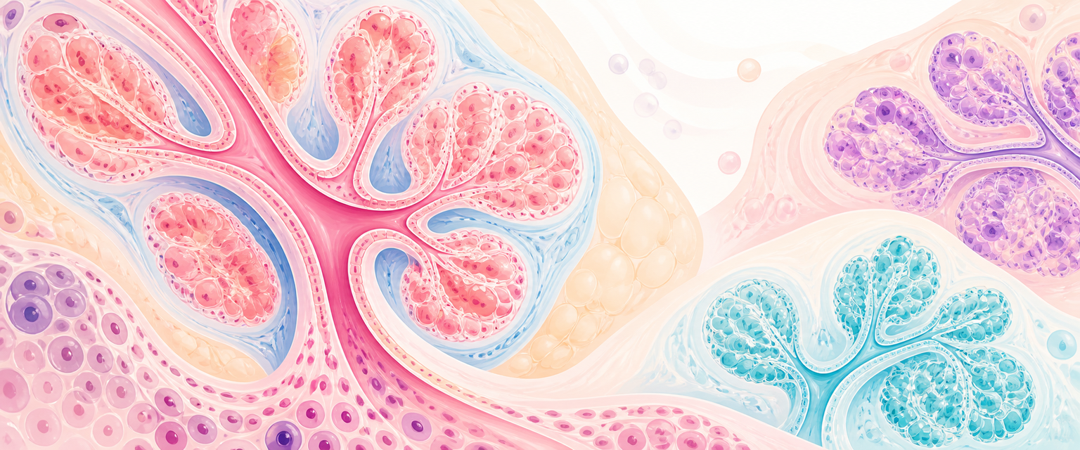
Female Breast
Start from the simplest true statement: the breast is a gland in the skin. More precisely, it is a modified apocrine sweat gland — a compound exocrine gland that, instead of secreting sweat onto the skin surface, has been repurposed to synthesise and deliver milk to an infant. Everything else in this chapter is an elaboration of that single idea. A gland needs secretory units (alveoli), a duct system to drain them (lactiferous ducts), a stroma to support them (fat and fibrous tissue), a blood supply to feed synthesis, a lymphatic system to clear the interstitium, and a control system (hormones and nerves) to switch secretion on and off. Build the breast in that order and the anatomy, histology and examination logic all follow from function.
The reason the breast matters so much in O&G is that, unlike most glands, it is not anatomically fixed. It is a dynamic, endocrine-responsive organ that is rebuilt repeatedly across a woman's reproductive life. Puberty, the menstrual cycle, pregnancy, lactation, contraception and menopause each remodel its ducts, lobules, stroma and vascularity. A Primary candidate must therefore be able to move from this structure to a clinical conclusion: a tender wedge in a lactating breast reflects duct-lobule drainage and inflammation; a new fixed mass in pregnancy is not dismissed as physiology; axillary nodes are examined because most lymph leaves the breast through the axilla; nipple stimulation drives hypothalamic-pituitary output through the prolactin and oxytocin reflexes.
This chapter ascends deliberately — from gross structure, through embryology and histology, to vascular, lymphatic and neural supply, and finally to the hormone-driven changes of pregnancy and lactation that make examination harder. It stays with structure, histology and examination logic. Detailed treatment of mastitis, puerperal sepsis and cancer in pregnancy belongs in the Intermediate and Final chapters linked below.
Core Knowledge
Surface Anatomy and Fascial Relationships
The adult female breast lies mainly over pectoralis major, with extension onto serratus anterior and the upper abdominal wall. In standard anatomical description it extends from the second to sixth ribs and from the lateral sternal edge to the mid-axillary line, but the clinically important point is that breast tissue is not limited to the visible mound. The axillary tail of Spence passes superolaterally through the deep fascia toward the axilla, so examination that stops at the apparent breast edge misses real breast tissue.
The breast is contained in the superficial fascia. It is not enclosed by a thick capsule. Fibrous septa, the suspensory ligaments of Cooper, run from skin to deep fascia through the gland. The posterior breast rests on the retromammary space, a loose areolar plane that allows the normal breast to move over pectoralis major. Loss of that mobility raises concern for deep fixation by inflammation, scarring or malignancy.
| Structure | Anatomical Description | O&G And Examination Relevance |
|---|---|---|
| Breast mound | Gland, fat and fibrous stroma in superficial fascia | Size and texture vary widely; asymmetry can be normal if long-standing |
| Axillary tail | Superolateral projection into axilla | Must be palpated in every breast examination |
| Retromammary space | Loose areolar tissue between breast and deep fascia | Allows normal mobility; fixation is abnormal |
| Cooper ligaments | Fibrous septa from skin to deep fascia | Shortening can cause skin dimpling |
| Inframammary fold | Skin/fascial crease at lower breast | Common site for intertrigo and examination blind spot |
| Nipple-areolar complex | Nipple, areola, smooth muscle, ducts and glands | Latch, discharge, inversion and Paget-like change are assessed here |
Embryology and Developmental Anatomy
Breast development begins along the embryonic mammary ridge, or milk line, which runs from axilla to groin. The mammary glands themselves arise as a collection of modified sweat glands at the cranial (pectoral) end of this ridge — the ductal and glandular epithelium derives from surface ectoderm, while the supporting connective tissue and fat come from the underlying mesenchyme. In humans most of the ridge regresses except in the pectoral region. Failure of regression can leave accessory (supernumerary) nipples or accessory breast tissue, classically along the milk line and especially in the axilla. Because accessory tissue responds to the same hormones as normal breast, it may enlarge, become painful or even lactate during pregnancy and the puerperium — a useful explanation for an axillary swelling that appears only in the peripartum period.
Before puberty, the female breast contains only a small ductal system embedded in fibrous tissue, with a formed areola and small nipple but essentially no alveoli. At puberty (thelarche, typically around 9 to 12 years), the gland matures under a layered hormonal programme. Oestrogen drives ductal elongation, while cyclic progesterone after ovulation supports lobuloalveolar maturation. Sitting behind these two reproductive steroids is a set of permissive (metabolic) hormones required for the gland to grow and respond at all — growth hormone, thyroid hormone, parathyroid hormone, cortisol and insulin. The visible increase in breast size at puberty is largely fat deposition in the stroma, not a measure of glandular or lactation capacity. This is a common counselling point: a small breast can lactate effectively if the glandular tissue, nipple anatomy and milk removal are adequate, because lactation depends on alveolar mass and milk transfer, not on the volume of fat.
| Life Stage | Dominant Anatomical Change | Practical Consequence |
|---|---|---|
| Childhood | Rudimentary ducts, minimal lobules | True breast masses are uncommon; premature thelarche is developmental, not adult breast disease |
| Puberty | Ductal growth, fat deposition, areolar enlargement | Transient asymmetry and tenderness are common |
| Ovulatory cycles | Cyclical epithelial and stromal oedema | Luteal tenderness and nodularity may vary with cycle |
| Pregnancy | Ductal and lobuloalveolar proliferation, vascularity | Dense, nodular, tender breast; examination becomes harder |
| Lactation | Secretory alveoli and active milk transport | Engorgement, milk stasis, mastitis and abscess anatomy become relevant |
| Menopause | Lobular involution, fat replacement | Glandular density falls, but cancer risk rises with age |