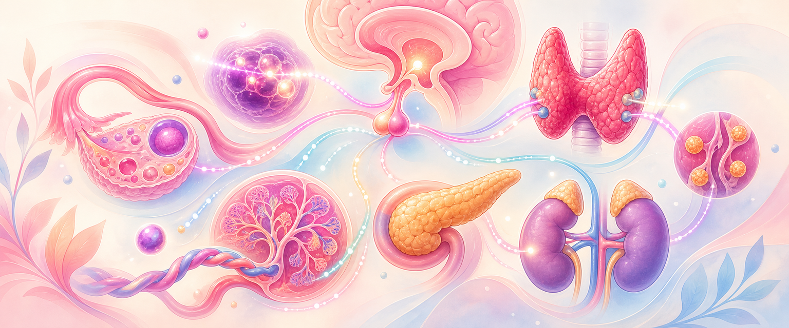
Endocrine Organs and Spinal Innervation
Start with the simplest idea the rest of the chapter rests on. A hormone is a chemical message released into the blood by one tissue that changes the behaviour of a distant tissue carrying the matching receptor. An endocrine gland is the organ that makes and releases it. Because the message travels in blood rather than down a duct, a tiny gland can change the whole body, and anything that alters how the message is carried, cleared or read will look like a glandular disorder even when the gland is normal. Hold that single sentence: it is the reason pregnancy can make a healthy thyroid or pancreas look abnormal on a blood test.
From that first principle, endocrine anatomy becomes the anatomy of control loops. The hypothalamus controls the pituitary through blood vessels and axons; the pituitary controls the ovary, thyroid, adrenal and breast; the target glands feed their hormones back to the brain to set the next command; and in pregnancy a placenta is added that changes binding proteins, makes its own hormones, alters metabolism and so rewrites how every result is interpreted. Each control loop has the same three parts — a command from above, a gland that obeys, and a feedback signal that returns — so once you can read one loop you can read them all.
Pelvic neuroanatomy is the partner idea: the anatomy of signal routes. Nerves are the body's fast, wired messaging system, the counterpart to the slow, broadcast endocrine system. In the pelvis these routes carry visceral pain from the uterus, somatic pain from the perineum, parasympathetic commands for bladder emptying, sympathetic tone, pudendal continence and — when they are cut or stretched — the deficits of surgical and obstetric nerve injury. Where the endocrine half asks which loop is broken, the neural half asks which route was interrupted.
For Primary FCOG, the aim is not to recite hormone lists. It is to use anatomy to solve O&G problems: why a pituitary mass affects vision and cycles; why a virilised 46,XX infant may have an adrenal enzyme problem; why thyroid enlargement changes airway assessment; why gestational diabetes is a stress test of beta-cell reserve; why pudendal block does not abolish contraction pain; and why pelvic surgery can injure autonomic, obturator or abdominal-wall nerves. Read the chapter in order: each section assumes only the one before it, building from what a gland is, through the master control loop, out to each gland, and finally to the nerves that share the same pelvis.
Core Knowledge
Organising Principles
Endocrine glands are ductless, vascular organs that release hormones into blood. Their arrangement differs from exocrine glands, where ducts carry secretions to an epithelial surface. Endocrine histology is therefore built around epithelial cords, follicles, nests or islets lying close to capillaries. This vascularity matters clinically: endocrine disorders often present systemically, small tumours can produce large biochemical effects, and pregnancy can change hormone transport without changing the gland itself.
| Principle | Anatomy Behind It | Clinical Use |
|---|---|---|
| Portal blood flow | Hypothalamic hormones reach anterior pituitary through portal vessels | Small hypothalamic signals control large pituitary outputs |
| Neurosecretion | Posterior pituitary releases hormones made in hypothalamic neurons | Oxytocin and vasopressin are stored, not made, in posterior pituitary |
| Feedback loops | Target hormones act on pituitary and hypothalamus | High TSH with low T4 means primary thyroid failure |
| Cell zonation | Different cell groups make different hormones | Adrenal cortex zones explain aldosterone, cortisol and androgen patterns |
| Shared embryology | Some organs have mixed tissue origins | Adrenal cortex and medulla behave like different organs |
| Neural root maps | Pain and motor pathways enter specific spinal levels | Labour analgesia and pudendal block are root-level problems |
A short note on embryology makes the later sections cohere rather than feel like a list of unrelated glands. The endocrine system is built from three different germ-layer sources, and that origin predicts behaviour. The anterior pituitary grows up from the roof of the developing mouth (an ectodermal pouch), so it is true glandular tissue. The posterior pituitary grows down from the floor of the brain as neural tissue, so it stores hormones made by neurons rather than manufacturing its own. The thyroid descends from the floor of the pharynx, dragging a track (the thyroglossal duct) that explains midline neck remnants. The parathyroids and part of the thymus arise from pharyngeal pouches and migrate, which is why parathyroid tissue can end up in unexpected (ectopic) positions. The adrenal cortex is mesoderm while the adrenal medulla is neural-crest tissue — effectively a sympathetic ganglion wrapped inside a steroid gland. You do not need to memorise the embryology for its own sake; you need it because it tells you why one organ behaves like two, why glands turn up in odd places, and why the posterior pituitary is a storage depot rather than a factory.
Hypothalamus and Pituitary
The hypothalamus lies in the floor and walls of the third ventricle. It integrates neural, metabolic, stress and reproductive signals and communicates with the pituitary through two anatomical routes. The anterior pituitary receives hypothalamic releasing or inhibiting hormones through the hypophyseal portal circulation. The posterior pituitary is neural tissue connected to hypothalamic nuclei by axons running through the infundibulum.
The pituitary sits in the sella turcica, beneath the optic chiasm and close to the cavernous sinuses. This relationship explains headache and bitemporal visual-field symptoms in pituitary enlargement. In pregnancy the lactotroph population expands, so the gland enlarges physiologically; marked symptoms or acute headache still require clinical escalation, but the anatomical reason for pregnancy sensitivity is glandular hyperplasia in a small bony fossa.
| Component | Anatomy/Histology | Hormones Or Signals | O&G Relevance |
|---|---|---|---|
| Hypothalamic GnRH neurons | Pulsatile neural secretion to portal vessels | GnRH pulses | Puberty, ovulation, hypothalamic amenorrhoea |
| Anterior pituitary gonadotrophs | Glandular cells | FSH, LH | Follicle development, ovulation, menopause interpretation |
| Anterior pituitary lactotrophs | Dopamine-inhibited cells | Prolactin | Galactorrhoea, anovulation, lactation |
| Anterior pituitary thyrotrophs | TRH-responsive cells | TSH | Thyroid assessment in fertility and pregnancy |
| Anterior pituitary corticotrophs | CRH-responsive cells | ACTH | Adrenal cortisol and androgen pathways |
| Posterior pituitary | Axon terminals from hypothalamus | Oxytocin, vasopressin | Milk ejection, uterine contraction physiology, water balance |
The most tested point is pulsatility. Pulsatile GnRH stimulates FSH and LH. Continuous GnRH exposure suppresses gonadotrophins after an initial flare. The same molecule can therefore stimulate or suppress the axis depending on delivery pattern. This is anatomy, not just pharmacology: portal-vessel exposure over time determines pituitary cell response.
Prolactin is the anterior pituitary exception. Most anterior pituitary axes are stimulated by hypothalamic releasing hormones; prolactin is mainly under tonic dopamine inhibition. Damage to the pituitary stalk can raise prolactin by removing dopamine restraint. Hyperprolactinaemia suppresses GnRH pulsatility, so the reproductive presentation may be oligomenorrhoea, amenorrhoea, infertility or galactorrhoea.
The posterior pituitary deserves its own paragraph because it works on a completely different principle from the anterior gland. It is not a factory — it is a storage and release terminal. Two hypothalamic nuclei, the supraoptic and paraventricular nuclei, manufacture oxytocin and vasopressin (antidiuretic hormone, ADH) in their cell bodies, then send the hormones down long axons through the infundibulum to be stored in the posterior lobe and released into the blood on demand. This is the meaning of "neurosecretion": the gland you can see on a scan is only the release end of a neuron whose body sits in the brain. Oxytocin underpins the milk-ejection reflex and is part of uterine contraction physiology; vasopressin defends water balance by promoting renal water reabsorption. Both are covered in depth in the relevant Intermediate and Final chapters, but the Primary point is anatomical — they are stored, not made, in the posterior pituitary, so a stalk lesion can interrupt their delivery while the anterior pituitary axes are still intact.