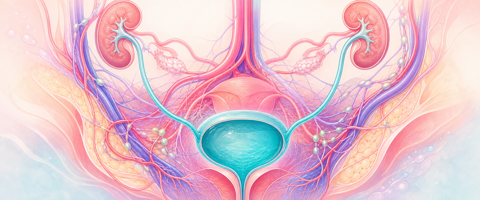
Why the Urinary Tract Belongs in an O&G Syllabus
Start with one idea: urine moves because of pressure gradients and because muscular walls actively push it. The kidneys generate urine, the renal pelvis and ureters propel it downhill by peristalsis, the bladder stores it at deliberately low pressure, and the urethra holds it in until the brain gives permission. Everything else in this chapter is a variation on that single pressure-and-flow system. Once you can see the tract as a plumbing circuit with active pumps and a controlled gate, the clinical disasters make sense rather than needing to be memorised. The vessels the ureter runs beneath, and the variants that bleed around it, are in Pelvic anatomy and supports.
The urinary tract is examined in Primary FCOG because it shares an operative and disease field with the uterus, cervix, vagina, adnexa and pelvic floor. A caesarean section mobilises bladder off the lower segment. A hysterectomy clamps tissue beside the ureter. An oophorectomy divides the infundibulopelvic ligament near the ureter at the pelvic brim. An anterior vaginal repair works immediately beneath bladder and urethra. Deep endometriosis and pelvic malignancy can encase the ureter. Prolonged obstructed labour can compress the bladder base and urethra until ischaemic fistula forms. You cannot operate safely in the female pelvis, nor interpret a pregnant woman's "back pain and fever", without knowing where this system runs and how it behaves.
Learn the tract as a sequence of jobs:
- kidneys filter blood, concentrate waste and form urine;
- renal pelvis and ureters conduct urine by active peristalsis along a retroperitoneal route;
- bladder stores urine at low pressure and empties by a coordinated detrusor contraction;
- urethra maintains continence through mucosal seal, sphincter tone and pelvic-floor support;
- autonomic and somatic nerves switch the system between storing and voiding;
- pelvic support tissues decide whether urethral closure pressure rises appropriately during a cough, after birth trauma, or with prolapse.
| Part | Anatomical principle | O&G consequence |
|---|---|---|
| Kidney | Retroperitoneal organ draining to renal pelvis | Pregnancy hydronephrosis, pyelonephritis, renal colic |
| Ureter | Long retroperitoneal tube crossing the pelvic brim and passing under the uterine artery | Hysterectomy, adnexal surgery and endometriosis injury |
| Bladder | Low-pressure reservoir anterior to uterus, cervix and upper vagina | Caesarean injury, fistula, retention, anterior prolapse |
| Trigone | Fixed smooth triangle between the ureteric orifices and the internal urethral orifice | Cystoscopy, ureteric jets, bladder-base injury |
| Urethra | Short outlet embedded in anterior vaginal-wall support | Stress incontinence, urethral trauma, catheterisation |
| S2–S4 parasympathetic | Detrusor contraction and the voiding reflex | Postpartum retention, pelvic autonomic injury |
| Pudendal nerve S2–S4 | External urethral sphincter and pelvic floor | Voluntary continence, perineal trauma, pudendal block |
How the Tract Is Built (and Why Development Explains Adult Relations)
The adult relations make far more sense if you know where the parts came from, so we build the tract before we walk through it. Two embryological lineages converge to make the female lower urinary tract, and that convergence is the reason a gynaecologist injures the ureter while operating on the uterus.
The kidneys and ureters arise from intermediate mesoderm, the nephrogenic cord running alongside the developing gonad and reproductive ducts. Three successive kidney systems form in sequence — the transient pronephros, then the mesonephros, and finally the metanephros, which is the definitive kidney. The metanephros is induced by a diverticulum off the mesonephric (wolffian) duct called the ureteric bud: the bud branches repeatedly to form the collecting system — collecting ducts, calyces, renal pelvis and the ureter itself — while the surrounding metanephric mesenchyme forms the nephrons. This shared origin with the reproductive ducts, and the fact that the metanephric kidney begins low in the pelvis and ascends, explains both congenital variants (pelvic kidney, horseshoe kidney, duplex collecting systems) and why the ureter remains a close neighbour of the genital tract for life.
The bladder and urethra come from a different source. The cloaca is divided by the urorectal septum into a dorsal anorectal canal and a ventral urogenital sinus. The urogenital sinus has three parts: an upper vesicourethral portion that becomes the bladder and is continuous superiorly with the allantois (later the median umbilical ligament/urachus); a middle pelvic portion that forms the female urethra; and a lower phallic portion that becomes the vaginal vestibule in the female. So the bladder and urethra are endodermal urogenital-sinus derivatives, whereas the ureter is a mesodermal ureteric-bud derivative — they meet only when the lower ends of the mesonephric ducts and ureteric buds are incorporated into the bladder base to form the trigone. That is why the trigone is anatomically and histologically distinct from the rest of the bladder, and why the ureteric orifices and internal urethral orifice sit at fixed points on it.
| Adult structure | Embryological origin | Clinical echo |
|---|---|---|
| Kidney + collecting system + ureter | Ureteric bud + metanephric mesenchyme | Duplex/ectopic ureter, pelvic/horseshoe kidney |
| Bladder + trigone | Vesicourethral urogenital sinus (trigone from absorbed duct ends) | Urachal remnants, trigonal sensitivity |
| Female urethra | Pelvic part of the urogenital sinus (endodermal) | Distal squamous lining, paraurethral (Skene) glands |
| Vaginal vestibule | Phallic/lower urogenital sinus | Shared embryology with distal urethra and vulva |
The Fetal Kidney and Amniotic Fluid
The fetal kidney is functional from around 12 weeks, although nephron formation continues until roughly 36 weeks; after that, growth is by nephron enlargement, not new nephrons. Before birth the kidney makes urine but has minimal filtration capacity, so the maternal–placental interface handles most waste clearance. Fetal renal blood flow is only about 5% of the combined cardiac output, and glomerular filtration rate at term is around 30% of the adult value. The urine produced is hypotonic to isotonic, and from the second trimester onward fetal urine is the dominant source of amniotic fluid.
This single physiological fact has large clinical reach. If the kidneys cannot make urine (bilateral renal agenesis, severe dysplasia, polycystic disease) or the urine cannot escape (posterior urethral valves, bilateral pelvi-ureteric junction obstruction), liquor falls and oligohydramnios follows, with the downstream sequence of pulmonary hypoplasia and limb deformation. After birth the kidney must suddenly concentrate urine and excrete nitrogenous waste on its own; much of the normal 5–10% loss of birth weight in the first week is diuresis from the still-immature kidney. GFR then climbs roughly threefold over the first weeks and reaches adult levels by about two years. For Primary, the take-home is that a normal liquor volume on ultrasound is indirect evidence of a working fetal urinary tract, and a low one demands you ask whether the fetus can make urine and whether it can get out.
Kidneys and the Renal Pelvis
With the tract built, we can now walk it from top to bottom. The kidneys lie retroperitoneally on the posterior abdominal wall, between about T12 and L3, with the right kidney slightly lower because the liver sits above it. The suprarenal (adrenal) glands cap their superomedial poles. Each kidney is wrapped in renal capsule, perirenal fat and renal fascia. The hilum is a vertical slit on the medial border transmitting, from anterior to posterior, the renal vein, renal artery and renal pelvis, along with lymphatics and sympathetic fibres. Internally, papillae project into about a dozen minor calyces; minor calyces drain into two or three major calyces, which converge on the renal pelvis. The pelvis narrows into the ureter at the pelvi-ureteric junction (PUJ), the first of the physiological narrowings where a stone can lodge.