Clinical overview
Pelvic organ prolapse (POP) is the descent of one or more of the pelvic organs — bladder, uterus, bowel, or vaginal vault — into or through the vagina, the consequence of failure of the connective-tissue, muscular and fascial supports of the pelvic floor. It is very common: the lifetime risk of surgery for prolapse or incontinence has historically been quoted at around 1 in 9–11 women, and prevalence rises steeply with age and parity. Although benign, prolapse can be deeply distressing — women describe a vaginal bulge or "something coming down", pelvic pressure or dragging that worsens through the day and on standing, and associated bladder, bowel and sexual dysfunction.
Because this objective centres on aetiology, classification and causes, the weight of this chapter is the why and the what — the support anatomy that fails, the compartments that prolapse, the standardised language (POP-Q) used to describe it, and the risk factors that overload or weaken the system — before a working account of assessment and management. Two framing points anchor everything that follows. First, prolapse is a disorder of support, not of the organ itself: the bladder behind a "cystocele" is normal; it is the anterior vaginal wall support that has given way, so the suffix "-cele" names the bulging vaginal wall, not a herniated organ. Second, prolapse, urinary incontinence (urinary-incontinence) and voiding difficulty/retention (urinary-retention) frequently coexist and interact — repairing one can unmask or relieve another. Surgical practice has been substantially reshaped by the national pause on transvaginal mesh, returning native-tissue repair to the centre of care. This chapter links to urinary-incontinence, urinary-retention, and climacteric-and-menopause (oestrogen and connective tissue).
Core knowledge
The support system that fails — DeLancey's three levels
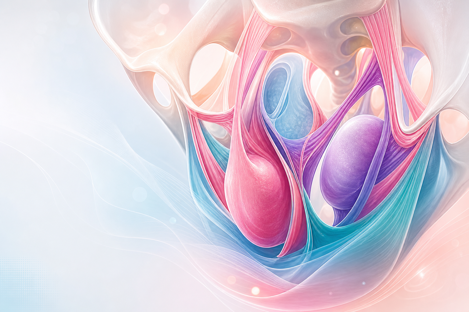
Figure C3.1 — How loss of support produces prolapse: DeLancey's three levels predict the compartment (apical/vault, cystocele, rectocele, enterocele as the true hernia), with levator + endopelvic fascia as the active and passive supports.
Pelvic support is provided by the levator ani muscles (the active, tonically contracted floor that keeps the urogenital hiatus closed) and the endopelvic fascia (the passive connective-tissue suspension). When the levator is damaged — classically by avulsion from the pubic ramus during vaginal birth — the hiatus widens and load is transferred to the fascia, which is not designed to bear it indefinitely; the muscular and fascial failures therefore compound one another. DeLancey described three levels of vaginal support, and the level that fails predicts the type of prolapse:
- Level I — apical suspension: the uterosacral and cardinal ligament complex suspends the cervix and upper vagina. Failure → uterine/apical prolapse and, after hysterectomy, vaginal vault prolapse and enterocele.
- Level II — lateral attachment: the mid-vagina is attached laterally to the arcus tendineus fascia pelvis ("white line") and the levator fascia. Failure → anterior (cystocele) and posterior (rectocele) wall prolapse.
- Level III — distal fusion: the lower vagina is fused to the perineal body, urethra and levators. Failure → urethrocele and perineal descent/deficiency.
Classification by compartment
Prolapse is described by the vaginal compartment that descends:
- Anterior compartment — cystocele (bladder), urethrocele (urethra), or cystourethrocele.
- Apical/middle compartment — uterine prolapse, or vaginal vault prolapse after hysterectomy; enterocele (a true peritoneal hernia containing small bowel, usually through the apex/posterior fornix — the only "-cele" that is a genuine hernia sac).
- Posterior compartment — rectocele (rectum) and perineal deficiency.
- Procidentia — complete eversion of the uterus and both vaginal walls outside the introitus.
Classification by severity — POP-Q

Figure C3.2 — POP-Q: the nine measured points (Aa, Ba, C, D, Ap, Bp, gh, pb, tvl) relative to the hymen (negative = above, positive = below) and the ordinal stages 0–IV.
The Pelvic Organ Prolapse Quantification (POP-Q) system (ICS/IUGA) is the international standard. It records the position, in centimetres, of defined vaginal points relative to the hymen — the fixed reference plane, where negative = above (proximal) and positive = below (distal) the hymen:
- Aa, Ba — two anterior-wall points; C — the cervix or vaginal cuff; D — the posterior fornix (omitted post-hysterectomy); Ap, Bp — two posterior-wall points; plus the measurements gh (genital hiatus), pb (perineal body) and tvl (total vaginal length).
- These condense into ordinal stages: Stage 0 (no descent), Stage I (leading edge >1 cm above the hymen), Stage II (leading edge within 1 cm above to 1 cm below the hymen), Stage III (>1 cm below the hymen but not complete), Stage IV (complete eversion/procidentia).
(The older Baden–Walker halfway system, grades 0–4, is still encountered.) Quantification matters because it gives reproducible, comparable descriptions across clinicians and over time, and it separates the leading compartment from the others.
Aetiology and causes — what overloads or weakens support
Prolapse develops when the load on the pelvic floor exceeds the strength of its supports. The causes group into what predisposes, incites, promotes and decompensates (the classic "four Ps"):
Predisposing / constitutional
- Connective-tissue quality — genetic predisposition (a positive family history is common); inherited collagen disorders (Ehlers–Danlos, Marfan) markedly increase risk; ethnic variation in prevalence (lower in some African and Asian populations, higher in white and Hispanic women).
Inciting — pregnancy and childbirth (the dominant modifiable cause)
- Vaginal delivery and increasing parity, macrosomia, prolonged second stage, instrumental delivery (instrumental-delivery), and levator-ani avulsion — pregnancy and vaginal birth are the single largest contributors, through both direct muscle/fascia injury and pudendal/levator denervation.
Promoting — chronically raised intra-abdominal pressure
- Obesity, chronic cough (smoking, COPD), chronic constipation with straining, heavy lifting/occupational load, and large pelvic masses or ascites.
Decompensating — loss of tissue integrity over time
- Ageing and oestrogen deficiency after the menopause — weaker, less vascular, less elastic connective tissue (see climacteric-and-menopause); previous pelvic surgery, especially hysterectomy (loss of apical/Level-I support predisposing to later vault prolapse); prior continence surgery (which can alter vaginal axis and provoke a new compartment); and neurological/denervation injury to the pelvic floor.
Symptoms by compartment
- General — a sensation of bulge/"something coming down", pelvic pressure or dragging worse on standing or straining, a palpable lump, and in procidentia ulceration, discharge and bleeding of exposed, keratinising mucosa.
- Anterior (cystocele) — urinary frequency, incomplete emptying, recurrent UTI, and the need to reduce the bulge to void (urinary-retention); stress incontinence may coexist or be "masked" by urethral kinking.
- Posterior (rectocele) — incomplete defecation, splinting/digitation (pressing on the vagina or perineum to empty the rectum), and straining.
- Apical — dragging, dyspareunia, coital difficulty, and (in procidentia) chronic discharge and ulceration.
Assessment
History
Bulge and pressure symptoms and their effect on daily life; urinary, bowel and sexual symptoms; obstetric and surgical history; the risk factors above; and crucially the woman's own goals — symptom relief, a wish to retain the uterus, sexual activity, and fitness for surgery.
Examination
- Examine at rest and on Valsalva, and standing if the findings do not match the symptoms, ideally with a Sims speculum, assessing each compartment and the apex systematically and quantifying with POP-Q.
- Assess pelvic-floor muscle contraction, genitourinary atrophy, and any mucosal ulceration.
- Perform a cough stress test and assess for occult (masked) stress incontinence after reducing the prolapse — a large cystocele can hide SUI that appears once the prolapse is repaired.
Investigations
Largely clinical. Urinalysis and post-void residual; urodynamics where incontinence coexists or before surgery (and to detect occult SUI); renal tract imaging in procidentia to exclude ureteric kinking and hydronephrosis.
Management

Figure C3.3 — The "four Ps" of why support fails (predisposing, inciting, promoting, decompensating) and the symptom-led management ladder (reassure → PFMT → vaginal oestrogen → pessary → native-tissue/apical surgery); transvaginal mesh = research only.
Matched to symptoms, severity, and the woman's wishes — asymptomatic prolapse needs no treatment beyond reassurance and risk-factor modification.
- Conservative — lifestyle (weight loss, treat constipation and chronic cough), supervised pelvic-floor muscle training (most effective in milder, Stage I–II prolapse), and vaginal oestrogen for atrophy.
- Pessaries — ring (with or without support) or shelf/Gellhorn devices; an excellent option for those who decline or are unfit for surgery, who have not completed childbearing, or who simply prefer to avoid an operation. They require periodic review (typically 4–6 monthly) and topical oestrogen to prevent erosion, ulceration and (rarely) fistula from neglect.
- Surgery — selected by compartment and apex:
- Anterior/posterior native-tissue repair (anterior/posterior colporrhaphy).
- Apical procedures — vaginal hysterectomy with apical suspension, sacrospinous fixation or uterosacral ligament suspension (vaginal routes), or sacrocolpopexy (abdominal/laparoscopic, the most durable for vault prolapse). Uterine-preserving hysteropexy is an option where the uterus is to be retained.
- Obliterative surgery (colpocleisis) — highly effective and low-morbidity for frail women who do not wish to retain coital function.
- Vaginal mesh for prolapse is restricted to research use only under the current national pause; native-tissue repair and abdominal sacrocolpopexy (which places mesh via a non-transvaginal route) are the mainstays. Counsel explicitly about the mesh history and the chosen approach.
Red flags / pitfalls
- Procidentia with ureteric obstruction/hydronephrosis — image the renal tract; high-grade prolapse can threaten the upper tracts.
- Ulcerated, bleeding exposed mucosa — distinguish a benign decubitus ulcer from malignancy; biopsy if atypical or non-healing after reduction and oestrogen.
- Missing masked stress incontinence — assess with the prolapse reduced; SUI appearing after an anterior repair is a common, avoidable disappointment.
- Operating without addressing the apex — unsupported apical descent is the commonest reason anterior repairs fail; always assess and support the apex.
- Counselling failures on mesh — women must understand the transvaginal-mesh restriction and the native-tissue/abdominal alternatives.
- Pessary neglect — an unreviewed pessary can erode, become impacted, or fistulate; schedule review and use vaginal oestrogen.
- Forgetting voiding dysfunction — a large prolapse may cause incomplete emptying or frank retention (urinary-retention); check residuals.
- Surgery on an unmodified risk profile — uncorrected obesity, cough or constipation predicts recurrence.
Evidence anchors
- NICE NG123 — Urinary incontinence and pelvic organ prolapse in women: management (2019, last reviewed March 2025) — assessment, conservative care, pessaries, surgical options, and the transvaginal-mesh restriction.
- ICS / IUGA — POP-Q standardised terminology and quantification of pelvic organ prolapse.
- RCOG Green-top Guideline 46 — Post-Hysterectomy Vault Prolapse; RCOG/BSUG prolapse-surgery guidance.
- Independent Medicines and Medical Devices Safety Review ("First Do No Harm", Cumberlege 2020) — basis of the vaginal-mesh restriction.
- South African EML / NDoH Standard Treatment Guidelines — pessary and topical-oestrogen provision.
