Clinical overview
A gynaecological fistula is an abnormal, epithelialised communication between the genital tract and an adjacent hollow viscus — most often the urinary tract (bladder, ureter, urethra) or the bowel (rectum, colon). The cardinal symptom is continuous, uncontrollable leakage of urine or faeces into the vagina which — unlike stress or urgency incontinence (urinary-incontinence) — is constant and unrelated to effort or urge. Beyond the physical morbidity (perineal excoriation, recurrent infection, stone formation, odour), fistulae are socially catastrophic: in the obstetric setting they cause profound stigma, isolation, loss of livelihood, divorce and destitution, which is why obstetric fistula is treated as a maternal-health and human-rights priority as much as a surgical problem. The global burden remains large — hundreds of thousands of women live with untreated obstetric fistula, overwhelmingly in low-resource settings, and it is a marker of inequitable access to emergency obstetric care.
Because this is a pathology/pathophysiology objective, the emphasis here is on how fistulae form, the tissue events that produce them, where they occur, and how they are classified — the foundation that explains both prevention and repair. The single most important conceptual divide is between the obstetric fistula (ischaemic-pressure necrosis from obstructed labour, the dominant cause in low-resource settings) and the iatrogenic/surgical fistula (sharp or thermal injury, the commoner cause in better-resourced settings, including much of urban South Africa). This chapter links to urinary-incontinence (the key differential), urinary-retention, and the obstetric prevention context of safe-caesarean-technique.
Core knowledge
Types by anatomical communication
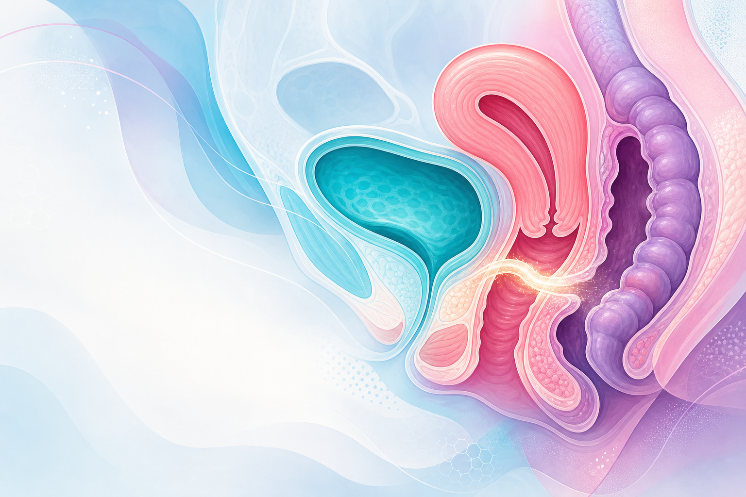
Figure C4.1 — The fistula map: types named by the organs they connect (vesicovaginal, ureterovaginal, urethrovaginal, vesicouterine/Youssef, rectovaginal), with the key discriminating features of each.
- Vesicovaginal fistula (VVF) — bladder to vagina; the commonest urogenital fistula; continuous urinary leakage per vaginam with, in larger fistulae, little or no normal voiding.
- Ureterovaginal fistula — ureter to vagina; classically continuous leakage with preserved normal voiding (the contralateral ureter still drains the bladder normally); almost always iatrogenic after pelvic surgery.
- Urethrovaginal fistula — urethra to vagina; leakage, spraying on voiding, or asymptomatic if proximal and small; if it involves the continence mechanism, repair may not restore continence.
- Vesicouterine fistula — bladder to uterus; a recognised complication of caesarean section, classically presenting with cyclical haematuria (menouria), amenorrhoea and urinary continence — the triad of Youssef syndrome.
- Rectovaginal fistula (RVF) — rectum to vagina; passage of flatus or faeces per vaginam; from obstetric anal-sphincter injury (oasis), obstructed labour, surgery, Crohn's disease, or malignancy.
- Complex/combined — large obstetric fistulae may involve bladder, urethra and rectum together, with extensive circumferential tissue loss.
Pathophysiology by cause

Figure C4.2 — The two mechanisms: obstetric ischaemic pressure necrosis (obstructed labour) vs iatrogenic sharp/thermal surgical injury — with their contrasting tissue quality, timing, and outcomes.
Obstetric — ischaemic pressure necrosis (the classic mechanism)
In prolonged, neglected obstructed labour, the fetal head is impacted against the maternal bony pelvis, compressing the intervening soft tissues — the bladder base, urethra, anterior vaginal wall and rectum — between the descending head and the pubic symphysis/sacrum. Sustained compression occludes the blood supply: ischaemia → pressure necrosis → sloughing of the devitalised tissue, and a fistula becomes apparent 3–10 days postpartum as the slough separates (so the leakage characteristically starts a few days after delivery, often after a stillbirth). Because the insult is ischaemic and widespread, the surrounding tissue is fibrotic, scarred, contracted and poorly vascularised, defects are frequently large and complex, and associated injuries cluster into the "obstructed labour injury complex": urethral loss and stress incontinence even after closure, vaginal stenosis, secondary infertility and amenorrhoea (Asherman-like or hypothalamic), and foot-drop from lumbosacral trunk/common peroneal compression. This is fundamentally a disease of delayed or absent emergency obstetric care — hence its concentration where skilled birth attendance and timely caesarean are unavailable, and its prevention through the partograph/WHO Labour Care Guide and timely caesarean (safe-caesarean-technique).
Iatrogenic / surgical — sharp or thermal injury
The commoner mechanism wherever obstetric care is good. A fistula follows direct (sharp) injury, a suture inadvertently placed through the bladder, devascularisation, or thermal/energy injury during pelvic surgery — most often total (especially abdominal) hysterectomy, producing the classic supratrigonal VVF at the vaginal vault, and caesarean section; ureteric injury during pelvic surgery produces a ureterovaginal fistula. These typically present 1–3 weeks post-operatively as the injured tissue breaks down and urine tracks to the vagina. The surrounding tissue is usually better vascularised than in obstetric fistula, the defects smaller and cleaner, and the repair prognosis correspondingly better.
Radiation
Pelvic radiotherapy (e.g. for cervical carcinoma) causes a progressive obliterative endarteritis → chronic ischaemia and fibrosis, which can generate fistulae months to years later. Radiation fistulae are notoriously difficult — poorly vascularised, slow to heal, prone to recurrence, and demanding tissue interposition at repair — and recurrent malignancy must always be excluded as the cause.
Malignancy and infection
Direct invasion by advanced cervical, vaginal or rectal cancer; and infective/inflammatory causes — lymphogranuloma venereum, genital tuberculosis, schistosomiasis, and Crohn's disease (a leading cause of RVF in high-income settings).
Classification
Standardised classification guides prognosis and surgical planning. The key descriptors in any system are site, size, involvement of the urethra/continence mechanism, degree of scarring/tissue loss, and ureteric involvement — these predict the chance of closure and the risk of residual (post-repair) incontinence.
- Goh classification (2004) — the most widely used; grades the fistula by the distance of its distal edge from the external urethral meatus (Types 1–4, where higher types lie progressively closer to or involve the urethra/continence mechanism), the size of the defect, and special considerations (scarring, circumferential loss, prior repair).
- Waaldijk classification — based on whether the continence (closure) mechanism and urethra are involved (Types I/II/III), which is directly predictive of post-repair continence.
The decisive differential
The pathophysiology yields a simple bedside rule: continuous leakage that is unrelated to effort or urge is a fistula until proven otherwise — and continuous leakage with otherwise normal voiding points specifically to a ureterovaginal fistula.
Assessment
History and examination
- Continuous leakage of urine and/or flatus/faeces per vaginam; the timing relative to delivery (obstetric: a few days postpartum) or surgery (iatrogenic: 1–3 weeks); obstetric history (obstructed/prolonged labour, stillbirth), surgical and oncological history, and radiotherapy.
- Examination — often best under anaesthesia — to locate the fistula, define site, size, scarring and the state of the urethra/continence mechanism, and exclude multiple tracts.
Investigations
- Dye tests — instil dilute methylene blue into the bladder to confirm a vesicovaginal tract. The three-swab (tampon) test localises and distinguishes types: a proximal swab stained blue indicates a vesicovaginal fistula, whereas a distal swab that is wet but not blue (clear urine) indicates a ureterovaginal fistula.
- Cystoscopy ± examination under anaesthesia; IV urography / CT urogram or retrograde studies to define ureteric involvement; renal function and imaging.
- Biopsy of the fistula edges where malignancy or radiation recurrence is possible.
Management

Figure C4.3 — Work-up and repair: the three-swab dye test to localise the tract, and the management ladder (prevent → catheter for fresh small fistulae → timed repair → definitive layered repair → tissue interposition → after-care); "don't squander the first repair".
The principles flow directly from the pathophysiology:
- Prevention is paramount. For obstetric fistula: skilled birth attendance, the partograph/WHO Labour Care Guide, and timely caesarean for obstructed labour. For iatrogenic fistula: meticulous surgical technique, ureteric identification, and immediate recognition and repair of intra-operative injury.
- Early / conservative management. A small, fresh fistula (especially iatrogenic) may close with continuous bladder catheter drainage for several weeks — keeping the bladder empty lets the tract heal — combined with skin care, nutrition, and treatment of infection.
- Surgical repair is the definitive treatment, by a trained fistula surgeon working with a holistic team. Timing allows inflammation and slough to settle (and, in obstetric fistula, the tissue to demarcate — traditionally a wait of around 6–12 weeks, though selected fresh, clean fistulae are repaired early). Principles: tension-free, layered closure of healthy tissue, with interposition of a well-vascularised flap (e.g. a Martius labial fat-pad graft, or omentum) in scarred, radiation or recurrent cases; the Latzko technique is used for vault VVF. The route (vaginal or abdominal) is dictated by site, access and complexity. First-attempt closure offers the best chance of success, so it should be done well by an experienced surgeon.
- Post-repair (residual) incontinence (POFRI) is an important recognised outcome even after anatomically successful closure — typically from urethral/sphincter involvement — and is assessed and managed along FIGO good-practice lines.
- Holistic care. Physical rehabilitation, treatment of associated injuries (foot-drop, vaginal stenosis), nutritional support, and psychosocial care and community reintegration — surgical closure alone does not undo the social harm.
Red flags / pitfalls
- Mistaking a fistula for ordinary incontinence — continuous leakage unrelated to effort or urge is a fistula until excluded (urinary-incontinence).
- Continuous leakage with normal voiding — points to a ureterovaginal fistula; do not stop the work-up at the bladder.
- Missing ureteric involvement — always assess the upper tracts before repair; a missed ureteric fistula dooms a bladder repair.
- Operating on inflamed or sloughing tissue too early — wait for demarcation/settling; premature repair on devitalised tissue fails.
- Forgetting malignancy/recurrence in radiation- or cancer-associated fistulae — biopsy the edges.
- Vesicouterine fistula presenting as cyclical haematuria/amenorrhoea (Youssef syndrome) after caesarean — easily overlooked.
- Squandering the first repair — the first attempt has the highest success rate; refer to an experienced fistula surgeon rather than attempt an under-resourced repair.
- Treating the hole and neglecting the woman — obstetric fistula carries nerve injury, stigma and destitution; closure is necessary but not sufficient.
Evidence anchors
- FIGO — good practice recommendations to standardise assessment of outcomes following vesicovaginal fistula surgery (Browning et al., Int J Gynecol Obstet, 2025) and FIGO expert opinion on management of post-obstetric fistula repair incontinence (POFRI) (Goh et al., 2025).
- FIGO Global Competency-Based Fistula Surgery Training Manual and the GFMER Obstetric Fistula modules (2024 update) — diagnosis, classification, management.
- Goh classification (2004) and Waaldijk classification of urogenital fistula.
- WHO — obstetric fistula prevention and the "Ending Fistula" campaign (skilled birth attendance, partograph/Labour Care Guide, timely caesarean).
- South African context — obstetric fistula is now uncommon given facility delivery, with iatrogenic (post-hysterectomy/caesarean) fistula relatively more prominent; obstetric fistula persists in under-served and migrant populations.
