Cell Biology for O&G
Cell biology is not a catalogue of organelles. It is the microscopic language behind implantation, placental transport, steroid responsiveness, myometrial contraction, infection, inflammation, cancer, drug action and cell injury. In an O&G stem, words such as "trophoblast invasion", "oxytocin receptor", "progesterone withdrawal", "HPV oncogene", "mitochondrial hypoxia", "transport across the placenta" and "protein synthesis" are all asking the same thing: can you move from cellular structure to clinical consequence?
The Cell as the Smallest Clinical Unit
A cell has a controlled border, a genome, factories for proteins and lipids, an energy system, a skeleton, a waste-disposal system and communication links to neighbouring cells. O&G tissues behave differently because their cells emphasise different parts of that machinery. A syncytiotrophoblast is built for exchange and endocrine secretion; a myometrial smooth-muscle cell is built for contraction; a ciliated tubal epithelial cell is built for coordinated movement; a squamous cervical epithelial cell is built for barrier function; a cancer cell has escaped normal control over proliferation, adhesion and death.
| Cell structure | First-principle role | O&G relevance |
|---|---|---|
| Plasma membrane | Barrier, receptor platform, transport surface | Placental nutrient transfer, oxytocin receptor signalling, drug movement |
| Nucleus | DNA storage and transcription control | Steroid-regulated endometrium, HPV oncogenesis, fetal genetic testing |
| Ribosomes and rough ER | Translation and folding of secreted or membrane proteins | Peptide hormones, receptors, extracellular-matrix proteins |
| Smooth ER | Lipid synthesis, steroid metabolism, calcium handling | Steroidogenesis, smooth-muscle contraction, detoxification |
| Golgi apparatus | Glycosylation, sorting and packaging | Secreted proteins, membrane receptor trafficking, mucus glycoproteins |
| Mitochondria | ATP production and death signalling | Hypoxia, sepsis, haemorrhage, oocyte energy reserve |
| Lysosome, proteasome, autophagy | Controlled degradation and recycling | Antigen processing, infection response, tissue remodelling |
| Cytoskeleton | Shape, polarity, migration, mitosis, contraction | Trophoblast invasion, cilia, myometrial contraction, cancer spread |
| Junctions and basement membrane | Adhesion, barrier and communication | Epithelial integrity, myometrial synchrony, distinction between in situ and invasive disease |
The useful exam question is: which cellular compartment is doing the work? Rapid events usually involve membrane receptors, ion channels and phosphorylation. Slower tissue changes usually involve nuclear transcription and protein synthesis. Hypoxic collapse starts with mitochondria and ATP. Invasion requires cytoskeleton, adhesion switching, proteases and breach of basement membrane.
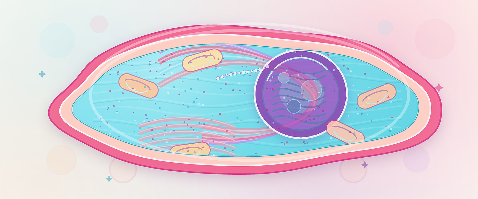
Figure A1.1 — The cell as the O&G engine: each organelle maps to a clinical consequence, so the exam move is to ask which compartment is doing the work.
Membrane Structure and Selective Transport
The phospholipid bilayer has hydrophilic heads facing aqueous fluid and hydrophobic tails facing inward. Cholesterol alters membrane fluidity. Membrane proteins act as channels, pumps, carriers, receptors, enzymes and adhesion molecules. The carbohydrate-rich glycocalyx contributes to recognition, immune interaction and epithelial surface behaviour.
The membrane is selective, not sealed. Small non-polar molecules such as oxygen, carbon dioxide and steroid hormones cross more easily. Charged solutes, glucose, amino acids, ions and most peptides require channels, carriers or active transport. The placenta is therefore not a passive sieve. It is a living epithelial transport organ with a large syncytiotrophoblast surface area, transporter expression, receptor-mediated uptake and metabolic activity.
| Transport mode | Energy requirement | Direction | O&G example |
|---|---|---|---|
| Simple diffusion | None | Down concentration gradient | Oxygen and carbon dioxide exchange |
| Facilitated diffusion | None | Down gradient through transporter/channel | Glucose movement through GLUT transporters |
| Primary active transport | Direct ATP | Against gradient | Na+/K+ ATPase maintaining electrochemical gradients |
| Secondary active transport | Uses another gradient | Coupled movement | Sodium-dependent nutrient transport |
| Endocytosis/exocytosis | ATP-dependent vesicular traffic | Bulk uptake or secretion | Hormone secretion, receptor recycling, immune-complex handling |
The clinical chain is straightforward:
| Cellular mechanism | Tissue effect | Clinical consequence |
|---|---|---|
| Transporter expression changes | Altered nutrient and drug transfer | Fetal growth, fetal exposure to medications |
| Membrane receptor upregulation | Greater responsiveness to ligand | Term myometrium becomes more contractile |
| Ion-channel opening | Calcium movement and electrical change | Smooth-muscle contraction or relaxation |
| Tight-junction disruption | Barrier failure | Ascending infection, epithelial inflammation |
| Membrane rupture after severe injury | Enzyme leakage and inflammation | Necrosis, sepsis-associated tissue damage |
Do not describe the placenta as "one membrane". Maternal blood bathes fetal-derived trophoblast; fetal capillaries lie within villi. Exchange depends on surface area, diffusion distance, transporter proteins, blood flow on both sides, and fetal-maternal concentration gradients.
Syncytiotrophoblast as a Cell Surface
For placental exchange, the key epithelial cell is the syncytiotrophoblast. Its maternal-facing microvillous membrane increases surface area in the intervillous space; its fetal-facing basal membrane is the last trophoblast barrier before villous stroma and fetal capillary endothelium. Nutrients, gases, antibodies and drugs therefore encounter cell membranes, transporters and vesicles before reaching the fetus.
| Substance/process | Dominant cellular route | O&G consequence |
|---|---|---|
| Oxygen and carbon dioxide | Diffusion driven by gradients and blood flow | Maternal hypoxia, anaemia or placental disease affects fetal oxygenation |
| Glucose | Facilitated diffusion through transporters | Maternal hyperglycaemia increases fetal substrate exposure |
| Amino acids and ions | Active and exchanger transport systems | Fetal concentrations can exceed maternal concentrations |
| Fatty acids | Enzymatic handling plus transport proteins | Placental metabolism shapes fetal lipid supply |
| IgG | Receptor-mediated vesicular transport | Maternal vaccination protects the neonate, especially when transfer time is adequate |
| Many drugs | Diffusion plus transporter/efflux modification | Fetal exposure is mechanism-specific, not simply "crosses/does not cross" |
This is the same membrane biology used in pharmacology. Small lipid-soluble non-ionised molecules cross more readily; charged, large or protein-bound molecules need transport or transfer poorly. Transporters and placental metabolism modify exposure but do not make the placenta a wall.

Figure A1.2 — Membrane structure and the five transport modes: small lipid-soluble molecules cross freely while charged, large or glucose loads need transporters — which is why the syncytiotrophoblast is a living transport organ, not a passive sieve.
DNA, RNA and Protein Synthesis
The central information flow is DNA -> RNA -> protein. DNA is stored in chromosomes in the nucleus. A gene is transcribed into pre-mRNA, introns are removed by splicing, and mature mRNA leaves the nucleus. Ribosomes translate mRNA codons into amino-acid sequence. Free ribosomes make cytosolic proteins. Ribosomes attached to rough endoplasmic reticulum make proteins destined for secretion, insertion into membranes or lysosomes. The Golgi apparatus then modifies, glycosylates, sorts and packages these proteins. Hold onto this sequence: the receptor signalling in the next section ultimately works by changing which of these proteins a cell makes.