Clinical overview
Genetics is not a niche academic subject in obstetrics and gynaecology — it sits at the centre of antenatal counselling, recurrent pregnancy loss, fetal medicine, sub-fertility, gynaecological oncology and paediatric gynaecology. Almost every working day a registrar fields a question that turns on a principle of inheritance: a couple who have had a baby with a neural tube defect and want to know their recurrence risk; a woman with a strong family history of breast and ovarian cancer asking whether her daughters are at risk; a pregnancy with a soft marker on the anomaly scan; a child with ambiguous genitalia (see ambiguous-genitalia); a primigravida whose brother has haemophilia. Answering these well requires you to reason from first principles rather than memorise condition lists, because the conditions are too many to memorise but the patterns of transmission are few.
In the South African context the burden and pattern of genetic disease has distinctive features. There is high consanguinity in some communities, founder mutations in others, a young population with high fertility, and — critically — limited and unevenly distributed access to clinical genetics services, karyotyping, chromosomal microarray and molecular testing. Most district and many regional hospitals have no on-site geneticist; the registrar is frequently the first and sometimes the only clinician to take a meaningful family history, recognise a pattern, and make a referral. Getting the basic principles right therefore has real consequences for which couples are offered prenatal diagnosis, carrier testing, or simply accurate reassurance.
Core knowledge
The molecular substrate
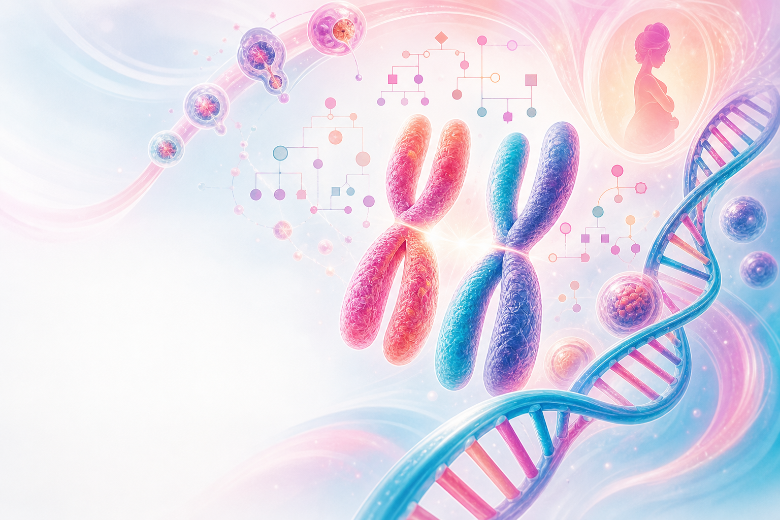
The human genome is carried on 46 chromosomes — 22 paired autosomes and two sex chromosomes (46,XX female; 46,XY male). Genes are segments of DNA that encode proteins; each gene occupies a fixed locus, and the alternative sequences at that locus are alleles. A person homozygous at a locus carries two identical alleles; a heterozygote carries two different ones. The observable trait is the phenotype; the underlying allelic make-up is the genotype. Reproductive cells (gametes) are haploid (23 chromosomes) and are produced by meiosis, in which homologous chromosomes pair, exchange material by recombination (crossing over), and segregate — the cellular basis of Mendel's laws and of genetic variability.
Figure K1.1 — From DNA to gametes: how chromosome structure, meiosis and fertilisation create genotype and phenotype.
Mendelian (single-gene) inheritance
Autosomal dominant — the phenotype is expressed in the heterozygote. An affected person typically has an affected parent (unless the variant is new), and each child of an affected person has, classically, a 1 in 2 (50%) chance of inheriting it, with males and females equally affected and male-to-male transmission possible. Dominant conditions often show variable expressivity (severity differs between carriers) and incomplete penetrance (some carriers never express the phenotype), which is why a pedigree may appear to "skip" a generation. Examples relevant to O&G include neurofibromatosis type 1, Marfan syndrome, myotonic dystrophy and the hereditary breast–ovarian cancer syndromes.
Autosomal recessive — the phenotype appears only in homozygotes (or compound heterozygotes). Affected children are usually born to unaffected carrier parents; if both parents are carriers, each pregnancy carries a 1 in 4 (25%) risk of an affected child, 1 in 2 of an unaffected carrier, and 1 in 4 of a homozygous-unaffected child. Consanguinity sharply increases the chance that both parents carry the same recessive allele and is therefore a key item of history. Cystic fibrosis, the haemoglobinopathies and many inborn errors of metabolism follow this pattern.
X-linked recessive — the variant lies on the X chromosome and is expressed in males (who are hemizygous — they have only one X) but usually not in heterozygous female carriers. A carrier mother transmits the variant to half her sons (who are affected) and half her daughters (who are carriers); an affected father transmits it to all his daughters (obligate carriers) but none of his sons, so there is no male-to-male transmission — a discriminating pedigree feature. Haemophilia A and B and Duchenne muscular dystrophy are the classic examples. X-linked dominant conditions (rarer) affect heterozygous females and may be lethal in males.
Beyond classical Mendelian patterns
Mitochondrial inheritance — mitochondria carry their own small circular genome and are transmitted almost exclusively through the oocyte cytoplasm, so mitochondrial disorders are passed from an affected mother to all her children, but never from the father. Heteroplasmy (a mixture of normal and mutant mitochondrial genomes within cells) explains the variable severity that characterises this group.
Genomic imprinting — for a minority of genes, only the maternally inherited or only the paternally inherited copy is expressed, the other being silenced. The phenotype therefore depends on the parent of origin. The textbook illustration is the deletion of the same chromosome 15q11–q13 region producing Prader–Willi syndrome when paternally derived and Angelman syndrome when maternally derived.
Multifactorial (polygenic) inheritance — most common congenital structural anomalies and adult diseases arise from the interaction of many genes of small effect with environmental factors. These do not follow Mendelian ratios; instead recurrence risk is empirical (derived from observed family data), typically in the order of a few per cent for first-degree relatives, and rises with the number of affected relatives and severity. Neural tube defects, congenital heart disease, cleft lip/palate and pyloric stenosis are standard examples — and the rationale for periconceptional folic acid.

Figure K1.2 — Inheritance pattern decoder: pedigree clues and recurrence-risk anchors for Mendelian and non-Mendelian patterns.
Chromosomal inheritance
Numerical errors (aneuploidy — e.g. trisomy 21, 18, 13; monosomy X) usually arise from meiotic non-disjunction and increase with maternal age. Structural rearrangements such as balanced translocations leave the carrier healthy but at risk of producing chromosomally unbalanced gametes, a recognised cause of recurrent miscarriage (see recurrent-pregnancy-loss). Aneuploidy is the bridge between these abstract principles and the everyday work of Down syndrome counselling (see down-syndrome-counselling).
Assessment
The three-generation pedigree
The single most useful and most under-used genetic "investigation" is a properly drawn three-generation family tree. Use standard symbols — squares for males, circles for females, a horizontal line for a couple, a vertical line to offspring, a diagonal slash for a deceased individual, a filled symbol for an affected individual and a dot for a known carrier. For each relative record age (or age and cause of death), affected status, miscarriages and stillbirths, and any infant or childhood deaths. Critically, ask directly about consanguinity ("are you and your partner related in any way, even distantly?") and about ethnic/community origin, because both shift the prior probability of recessive disease.
A well-constructed pedigree often reveals the inheritance pattern before any laboratory test: affected individuals in every generation with male-to-male transmission suggest autosomal dominant; affected males connected only through unaffected females, with no father-to-son transmission, suggest X-linked recessive; isolated affected children of unaffected (perhaps related) parents suggest autosomal recessive; transmission only through mothers suggests a mitochondrial disorder.
Targeted history and examination
Beyond the pedigree, ask about the index pregnancy or patient: maternal age, periconceptional folate, teratogen and alcohol exposure, maternal diabetes and epilepsy, and previous affected pregnancies. Examination of an affected child or fetus for dysmorphic features, growth and congenital anomalies helps direct testing, but pattern-recognition dysmorphology is a specialist skill — the registrar's job is to document carefully (with consent, photographs and measurements) and refer.
Investigations
Choose the test to match the question:
- Karyotype (chromosomal analysis) — detects aneuploidy and large structural rearrangements such as balanced translocations; still the test of choice when a balanced rearrangement is suspected.
- Chromosomal microarray (CMA) — higher-resolution detection of copy-number changes (microdeletions/duplications) but does not detect balanced rearrangements; increasingly first-line for fetal structural anomalies and for unexplained intellectual disability internationally, though availability in the SA public sector is limited and often restricted to tertiary referral.
- Targeted molecular/gene testing — for a specific suspected single-gene disorder or a known familial variant.
- Prenatal screening and diagnosis — aneuploidy screening uses the combined first-trimester test (nuchal translucency with βhCG and PAPP-A) and cell-free DNA (NIPT), which are screening tests; a positive screen is confirmed by diagnostic testing — chorionic villus sampling (from around the late first trimester) or amniocentesis (from the mid-second trimester), each carrying a small procedure-related miscarriage risk. These principles underpin the practical chapter on down-syndrome-counselling.
In South Africa, the National Health Laboratory Service (NHLS) provides cytogenetics and selected molecular testing through a small number of academic centres; access, turnaround time and cost frequently constrain what can be offered, and this reality must be discussed honestly with families rather than implying that every test available internationally is available locally.

Figure K1.3 — Pedigree to plan: three-generation history, pattern recognition, test selection and non-directive counselling.
Management
Genetic counselling: the core skill
The "management" of inheritance is overwhelmingly counselling, which must be non-directive, accurate and supportive. The accepted framework is: confirm the diagnosis where possible; establish the mode of inheritance; calculate the recurrence risk; explain the natural history and available options in clear language; and support the couple in reaching their own decision. Translate risks into both proportions and natural frequencies ("one in four, that is, 25 in every 100 pregnancies"), check understanding, and respect that the same numerical risk is acceptable to one couple and unacceptable to another. Counselling should ideally be pre-conceptional where a risk is known, because the widest range of options is then available.
Reproductive options to lay out
- Natural conception with prenatal diagnosis (CVS or amniocentesis) and the option of continuing or — within the legal framework — terminating an affected pregnancy.
- Termination of pregnancy for a serious fetal abnormality, governed in South Africa by the Choice on Termination of Pregnancy Act 92 of 1996 (see termination-of-pregnancy and sa-og-law); counsel within that legal framework.
- Preimplantation genetic testing with IVF, donor gametes, adoption, or a decision not to have further children — all to be presented neutrally where applicable, acknowledging that PGT and IVF are largely private-sector and cost-prohibitive for most South Africans.
- Carrier and cascade testing of at-risk relatives once a familial variant is identified, with attention to consent and confidentiality.
Prevention at population level
Some inherited disease is preventable or mitigable. Periconceptional folic acid reduces the recurrence and occurrence of neural tube defects, and South Africa's mandatory fortification of maize meal and wheat flour with folic acid is a population-level prevention programme; women with a previous affected pregnancy or on anti-epileptic drugs need a higher supplemental dose, which should be confirmed against the current SA guideline rather than assumed. Optimising maternal glycaemic control before conception and reviewing teratogenic medication are equally part of preconception genetic care.
Emergency considerations
Principles of inheritance are not in themselves an emergency, but they intersect with two situations that are. First, a neonate with ambiguous genitalia must be treated as a potential salt-wasting adrenal crisis from congenital adrenal hyperplasia until excluded — this is a neonatal emergency requiring urgent electrolytes and specialist input (see ambiguous-genitalia); do not assign or register a sex hastily. Second, in a known X-linked or recessive condition with a serious bleeding or metabolic phenotype (for example a male fetus at risk of severe haemophilia), the delivery plan, neonatal team and blood-product availability must be arranged in advance so that the birth is not the moment the diagnosis is first considered. The recurring safety message is the same: identify the genetic risk antenatally, communicate it clearly to the neonatal team, and plan the delivery setting accordingly rather than improvising at the bedside.
Red flags / pitfalls
- Treating screening as diagnosis. A high-chance combined test or NIPT result is not a diagnosis; it must be confirmed by CVS or amniocentesis before any irreversible decision. Conflating the two is a recurrent and serious error.
- Forgetting incomplete penetrance and variable expressivity. An apparently "skipped" generation does not exclude autosomal dominant inheritance; an unaffected obligate carrier can transmit a severe phenotype.
- Missing consanguinity and ethnicity. Failing to ask shifts recessive-disease risk estimates badly and is one of the commonest history omissions.
- Misreading new (de novo) variants. A negative family history does not exclude a dominant or X-linked condition — many arise de novo, and the recurrence risk for future pregnancies, though usually low, is not zero because of gonadal mosaicism.
- Applying Mendelian ratios to multifactorial conditions. Neural tube and cardiac defects do not recur at 25% or 50%; use empirical risks and do not frighten couples with the wrong model.
- Directive counselling. Steering a couple towards termination or continuation breaches the non-directive standard and, in SA, the autonomy protections of the National Health Act and the Choice on Termination of Pregnancy Act.
- Over-promising access. Offering tests (CMA, PGT, NIPT) that are not realistically available or affordable in the patient's setting sets up false expectations; be honest about NHLS turnaround, cost and referral pathways.
- Neglecting the neonatal emergency overlap. Ambiguous genitalia is CAH until proven otherwise; do not delay electrolytes or specialist referral while debating nomenclature.
Evidence anchors
- South African genetic and prenatal screening practice is framed by the National Integrated Maternal and Perinatal Care Guidelines for South Africa (NDoH, 2024), which sets out booking-visit risk assessment, the offer of aneuploidy screening and referral pathways within the SA level-of-care system.
- Aneuploidy screening principles — combined first-trimester test (NT + βhCG + PAPP-A), cell-free DNA / NIPT, and diagnostic CVS/amniocentesis, together with the Mendelian, X-linked, mitochondrial, multifactorial and imprinting patterns summarised here — follow the standard genetics teaching captured under the Genetics heading of the verified-sources reference; specific numerical screening performance and procedure-related loss rates should be quoted from the current local protocol before being given to a patient.
- Termination of pregnancy for fetal abnormality is governed by the Choice on Termination of Pregnancy Act 92 of 1996 (Act 92 of 1996, amended 2008), with consent and confidentiality framed by the National Health Act 61 of 2003 and HPCSA ethical guidance.
- Practical application of these principles to a single common aneuploidy is developed in the companion chapter on Down syndrome counselling.
Author's note on hedged facts: the Mendelian recurrence proportions (50%, 25%), the X-linked transmission pattern, mitochondrial maternal transmission and imprinting examples are standard textbook genetics, stated as such; they are not line-itemed with a specific guideline citation in VERIFIED-SOURCES.md and are therefore presented as standard teaching rather than attached to a fabricated reference. Procedure-related miscarriage rates and the exact higher folic-acid dose are deliberately not given as numbers here and must be confirmed against the current SA guideline before quoting to a patient.
