Clinical overview
Cervical cancer is the one common solid malignancy of women that we understand almost completely, from the first molecular insult to the invasive tumour — and it is the one we can almost completely prevent. That paradox is the heart of why South Africa carries one of the heaviest cervical cancer burdens in the world while the disease is being eliminated in countries with mature vaccination and screening. For the FCOG(SA) registrar, "discuss carcinogenesis" is not a request for a list of risk factors; it is a request to reason from a single virus, through a defined precursor lesion, to invasion — and to use that mechanistic chain to justify every preventive and therapeutic intervention you will offer.
The chain is deceptively simple: persistent infection of the cervical transformation zone with a high-risk human papillomavirus (HPV), integration of the viral genome with dysregulated expression of two oncoproteins (E6 and E7), progressive disruption of the host cell cycle and genome, and the emergence of a high-grade squamous intraepithelial lesion (HSIL) that, if unchecked, breaches the basement membrane. Each step is a clock that ticks over years to decades, and each step is a place to intervene — which is exactly why the WHO 90-70-90 elimination strategy targets vaccination (prevent infection), screening (detect the precursor), and treatment (excise the precursor or the early cancer) at three different points on the same curve. In a South African population with very high HIV prevalence, that clock runs faster and the precursor stage is more crowded, which reshapes how we screen and how aggressively we treat. This chapter builds the mechanism so the later staging and treatment objectives have foundations. See hpv-pathology and cin-pathophysiology for the lesion-level detail, and cervical-screening-sa and cin-management for the preventive programme this biology justifies.
Core knowledge
The transformation zone — where it all happens
Cervical carcinogenesis is anatomically anchored. The ectocervix is lined by non-keratinising stratified squamous epithelium; the endocervical canal is lined by simple columnar (glandular) epithelium. Between them lies the squamocolumnar junction, and because that junction migrates over a woman's life (everted onto the ectocervix at menarche and in pregnancy under oestrogen, then retreating into the canal after the menopause), the intervening band of metaplastic squamous epithelium — the transformation zone — is created by active metaplasia. It is the immature, dividing metaplastic cells of the transformation zone, not mature squamous or glandular cells, that are uniquely vulnerable to HPV-driven transformation. This is why almost all cervical squamous cancers and their precursors arise in the transformation zone, why colposcopy concentrates on it, and why an excisional treatment such as LLETZ aims to remove the whole zone. Knowing this anatomy explains the screening test (you must sample the zone) and the treatment (colposcopy localises it). See genital-anatomy.
HPV — the necessary cause
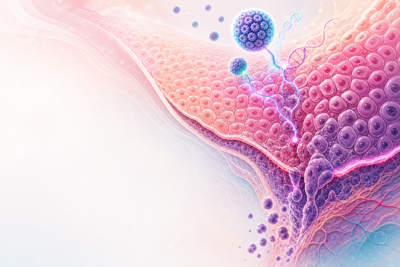
Figure D9.1 — The cervical carcinogenesis cascade: transient HPV → persistence → HSIL precancer → microinvasion → invasive carcinoma over ~10–20 years, with the three chances to interrupt it.
HPV is a small, non-enveloped, double-stranded circular DNA virus. More than 200 genotypes exist; about 14 are oncogenic ("high-risk", hrHPV), with HPV-16 and HPV-18 alone causing roughly 70% of cervical cancers worldwide. HPV-16 dominates squamous carcinoma; HPV-18 is over-represented in adenocarcinoma. Persistent infection with a high-risk type is a necessary cause — virtually no cervical cancer occurs without it — which is the single most important concept in this objective: it transforms cervical cancer from an idiopathic malignancy into an infectious disease with a precursor, and that is what makes vaccination and a molecular screening test rational.
The virus infects basal keratinocytes exposed through micro-abrasions in the transformation-zone epithelium. Most infections are transient: 80–90% clear within 1–2 years through cell-mediated immunity. It is persistence — the same high-risk type detectable over repeated tests — that defines risk. The viral life cycle is normally tied to keratinocyte differentiation, with the genome maintained as a low-copy episome.
E6 and E7 — the two oncoproteins

Figure D9.2 — The molecular engine: E6/E7 drive genomic instability, immortalisation (hTERT) and loss of cell-cycle control after integration disrupts E2.
Transformation is driven by sustained over-expression of two early viral genes, E6 and E7. In the WHO 2020 framework these map directly onto the histological grades:
- E7 binds and degrades retinoblastoma protein (pRb). pRb normally restrains the cell cycle by sequestering the E2F transcription factor at the G1/S checkpoint. Inactivating pRb releases E2F, forcing the cell into S-phase and continuous proliferation. A diagnostically useful consequence is over-expression of p16^INK4a — the cell raises p16 in a futile attempt to brake the cycle, so strong, block-positive p16 immunohistochemistry is a surrogate marker of high-risk HPV transcriptional activity and is used to confirm HSIL.
- E6 binds and targets p53 for proteasomal degradation (via the E6AP ubiquitin ligase). p53 is the guardian of the genome: it triggers cell-cycle arrest and apoptosis in response to DNA damage. Removing p53 disables apoptosis, so a cell that should die instead survives and divides. E6 also activates telomerase (hTERT), conferring replicative immortality.
Together, E6 and E7 abolish two independent tumour-suppressor checkpoints — uncontrolled proliferation plus loss of apoptotic surveillance — producing a cell that divides indefinitely, accumulates mutations, and cannot self-destruct. This is the molecular engine of hpv-pathology.
Integration and genomic instability
In the episomal state, E6/E7 are held in check by the viral E2 repressor protein. The pivotal event in progression is integration of the viral genome into the host chromosome, which typically disrupts the E2 open reading frame. Loss of E2 releases the brake on E6/E7, so their expression rises and becomes constitutive. Integration also induces host genomic instability — aneuploidy, copy-number changes, and the accumulation of secondary host mutations (for example in PIK3CA) — that drive the final transition to invasion. This is why integration broadly correlates with higher-grade disease, although integration is neither strictly required for nor exclusive to cancer. The take-home is the sequence: persistent productive infection → deregulated E6/E7 → genomic instability → invasion.
The precursor lesion — WHO 2020 / LAST terminology
Histopathology now uses the two-tier squamous intraepithelial lesion (SIL) terminology from the LAST project, adopted in the WHO Classification of Tumours of the Female Genital Tract, 5th edition (2020), replacing the older three-tier CIN system. Registrars must hold both, because clinical guidelines and the lab speak slightly different dialects:
| WHO 2020 / LAST | CIN equivalent | Biology |
|---|---|---|
| LSIL | CIN1 | Productive HPV infection; lower-third dysplasia; mostly regresses |
| HSIL | CIN2 and CIN3 | Transforming infection; true cancer precursor; treat |
LSIL reflects active viral replication and koilocytosis with abnormal cells confined to the lower third of the epithelium; the great majority regress spontaneously, especially in young women, and it is managed conservatively. HSIL reflects a transforming infection with disordered, immature cells occupying the middle/upper thirds (CIN2) or full thickness (CIN3); it is the genuine precursor and is treated. CIN2 is biologically heterogeneous — a mix of regressing LSIL-like and progressing HSIL-like lesions — which is why p16 immunostaining is used to stratify it and why conservative surveillance of CIN2 is reasonable in young women wishing to preserve fertility. Glandular precursors (adenocarcinoma in situ) follow the same HPV-driven logic but are harder to detect cytologically and are more often HPV-18 related. See cin-pathophysiology for full lesion detail.
Natural history and timelines
The single most exam-relevant fact about timelines is that progression is slow and stepwise. Most hrHPV infections clear; a minority persist; of persistent infections a minority produce HSIL; and untreated HSIL progresses to invasive cancer over a median of roughly 10–20 years. This long latency — typically a decade or more from persistent infection to invasion — is precisely what makes a screening programme with screening intervals of several years viable, and what makes any single missed screen recoverable. It is also why a cancer arising in a young woman, or rapid progression, should prompt thought about immunosuppression.
Cofactors — what tilts persistence toward progression

Figure D9.3 — How HIV/immunosuppression accelerates the HPV clock — faster persistence and progression, and the South African implications.
HPV is necessary but not sufficient; cofactors modulate whether a persistent infection progresses:
- HIV co-infection — the dominant cofactor in South Africa. HIV-associated CD4 depletion impairs the cell-mediated clearance of HPV, so women living with HIV have higher HPV prevalence, more multi-type and persistent infection, higher rates of HSIL, faster progression, higher recurrence after treatment, and a younger age at cancer. Invasive cervical cancer is an AIDS-defining illness. This biology is the reason SA screens women living with HIV earlier and more often (below). See hiv-gynaecology and hiv-counselling.
- Smoking — tobacco carcinogens are concentrated in cervical mucus and act as a co-mutagen, roughly doubling risk.
- Long-term combined oral contraceptive use (≥5 years) — a modest, partially reversible association; never a reason to withhold contraception, but relevant to counselling. See contraceptive-modalities.
- High parity, early coitarche, and other sexually transmitted infections (notably chlamydia, herpes) — markers of exposure and possible chronic inflammation.
- Immunosuppression generally (transplant, prolonged steroids) accelerates the same pathway.
Assessment

Figure D9.4 — Breach of the basement membrane = invasion: FIGO IA microinvasion (≤5 mm), lymphovascular space invasion, and routes of spread to pelvic/para-aortic nodes (FIGO IIIC).
"Assessing carcinogenesis" clinically means catching the process at the latest point that is still curable without major morbidity — ideally at the HSIL precursor stage. The diagnostic ladder maps onto the mechanism: detect the cause (HPV), find the lesion (cytology/colposcopy), and grade it (histology).
HPV testing detects the necessary cause and is the most sensitive primary screen. A high-risk HPV DNA/mRNA test, often with partial genotyping for the highest-risk types 16/18, identifies women carrying the transforming agent. Its high negative predictive value is what permits long screening intervals — a negative high-performance HPV test reassures for years. South Africa is moving toward HPV-DNA primary screening, informed by the South African DiaVACCS trial, with triage of HPV-positive women (genotyping, reflex cytology, or visual inspection with acetic acid, VIA) to decide who needs colposcopy or immediate treatment.
Cytology (Pap smear / liquid-based) detects the cellular consequence — dyskaryosis reflecting LSIL/HSIL. It is specific but less sensitive than HPV testing for a single test, and it depends on adequate transformation-zone sampling.
Colposcopy localises and characterises the lesion using acetowhitening (abnormal, high-nuclear-density epithelium turns white with 3–5% acetic acid), abnormal vascular patterns (punctation, mosaicism, atypical vessels), and Lugol's iodine (HSIL is glycogen-poor and stains non-uniformly). It directs biopsy. See colposcopy.
Histology is definitive and applies the WHO 2020 grading above, with p16 immunohistochemistry to confirm a transforming HPV infection (block-positive = HSIL) and to resolve ambiguous CIN2.
In suspected invasive disease, assessment shifts to staging — clinical examination, biopsy confirming stromal invasion, and imaging — under FIGO 2018, which (unlike its predecessor) permits imaging and pathology to assign stage, defines microinvasion (stage IA) by depth of invasion (≤5 mm) regardless of width, sizes IB by tumour diameter, and importantly classifies nodal disease as stage IIIC. MRI and transvaginal ultrasound assess local extent; PET-CT assesses nodes and distant metastasis. See endometrial-carcinoma for the contrasting FIGO 2023 molecular staging logic in that organ.
Management
Management here is the practical exploitation of the mechanism: interrupt the chain at vaccination, at the precursor, or at the earliest invasive disease. Treatment of established invasive cancer is covered with staging in other objectives; the carcinogenesis chapter's management remit is prevention and precursor treatment.
Primary prevention — vaccination (stop the infection)
HPV vaccines are prophylactic virus-like-particle vaccines that prevent acquisition of the targeted types; they do not treat existing infection. The nonavalent (9-valent) vaccine offers the broadest cover. The WHO Cervical Cancer Elimination Strategy (2020), with its 90-70-90 targets for 2030, places vaccination first: 90% of girls fully vaccinated by age 15. WHO (2022, with SAGE/RITAG support in 2023) endorsed a single-dose schedule, a pragmatic shift that has been adopted across Africa and dramatically simplifies programme delivery. South Africa has run a school-based HPV vaccination programme since 2014 for girls aged 9–14, now extending toward single-dose delivery and private schools. Vaccinating before coitarche maximises benefit because the vaccine cannot clear an infection already present.
Secondary prevention — screen and treat the precursor
This is the heart of the South African strategy and the most examinable SA-context content:
- SASOG / BetterGyn Clinical Guideline (2024) and the NDoH National Cervical Cancer Screening / Prevention & Control Policy define national practice. SA is transitioning to HPV-DNA primary screening; the historic public-sector cytology policy offered women three smears at 10-yearly intervals from age 30 (screening roughly ages 30–50).
- Women living with HIV are screened differently because of the accelerated biology above: screen at the time of HIV diagnosis and more frequently thereafter (approximately 3-yearly), regardless of age. This is the single most important SA divergence to quote in an exam.
- The screen-triage-treat pathway: HPV test → triage of positives (genotyping 16/18, reflex cytology, or VIA) → treatment of the precursor by thermal ablation (for ablation-eligible lesions) or large loop excision of the transformation zone (LLETZ) for HSIL or where excision/histology is needed. WHO's "screen-and-treat" or "screen-triage-treat" approach can compress this into fewer visits to reduce loss to follow-up — critical in a resource-constrained setting with a high default rate. See cin-management and cervical-screening-sa.
Treatment of the precursor is genuinely cancer-preventing: excising or ablating the transformation zone removes the deregulated-E6/E7 cells before they can complete the transition to invasion. Confirm test availability against the NHLS (the national laboratory that processes cytology and HPV testing) and the EML for the relevant consumables; access and turnaround are real-world rate limiters in SA.
Tertiary level — when invasion has occurred
Once the basement membrane is breached, management follows the ESGO/ESTRO/ESP cervical cancer guidelines (2023) and FIGO 2018 stage. Early disease is treated surgically (radical hysterectomy with nodal assessment, or fertility-sparing trachelectomy in selected small tumours). Locally advanced disease is treated with definitive concurrent chemoradiation — weekly cisplatin — plus image-guided adaptive brachytherapy, with the EMBRACE-II / GEC-ESTRO (ESTRO) standards defining modern brachytherapy. Immunotherapy has now entered first-line care: KEYNOTE-A18 added pembrolizumab to concurrent chemoradiation for high-risk locally advanced disease (FDA approved 12 January 2024, regardless of PD-L1), and KEYNOTE-826 established pembrolizumab plus chemotherapy ± bevacizumab first-line for recurrent/metastatic disease (PD-L1 CPS ≥1), with cemiplimab in the second-line recurrent setting. These are mechanism-consistent: an HPV-driven tumour expressing viral neoantigens is a rational immunotherapy target.
Red flags / pitfalls
- "HPV-positive equals cancer." It does not. Most infections clear; it is persistent high-risk infection that matters. Counsel accordingly to avoid harm and over-treatment, and never silently reinterpret a transient infection as disease.
- Over-treating LSIL/CIN1. LSIL is a productive infection that mostly regresses; ablating or excising every CIN1 causes obstetric harm (LLETZ raises preterm-birth risk) without preventing cancer. Treat HSIL; surveil LSIL.
- Forgetting the HIV-modified pathway. In SA the commonest examiner trap is applying the general-population screening age and interval to a woman living with HIV. She is screened at HIV diagnosis and more frequently regardless of age. A young woman with rapidly progressive or recurrent high-grade disease should prompt an HIV test.
- Sampling the wrong epithelium. A smear that misses the transformation zone is falsely reassuring; an inadequate or unsatisfactory result is not a normal result. After menopause the zone retreats into the canal, raising the false-negative risk.
- Confusing the terminologies. Know that LSIL=CIN1 and HSIL=CIN2/3; do not let a two-tier cytology report and a three-tier historical guideline desynchronise your management.
- Adenocarcinoma in situ is sneaky. Glandular precursors are HPV-18-biased, lie higher in the canal, and are poorly detected by cytology and ablation — they need excision and careful margin assessment.
- Assuming vaccination treats existing disease. The vaccine is prophylactic; a vaccinated woman still needs screening, and an infected woman is not cured by vaccinating.
Evidence anchors
- South African cervical cancer screening — SASOG / BetterGyn Clinical Guideline (2024) and the NDoH National Cervical Cancer Screening / Prevention & Control Policy — SA source of truth; transition to HPV-DNA primary screening (informed by the DiaVACCS trial); historic cytology policy (ages ~30–50, three smears 10-yearly); women living with HIV screened at HIV diagnosis and ~3-yearly regardless of age. Confirm exact intervals/algorithm against the current PDF.
- WHO Cervical Cancer Elimination Strategy (2020) — 90-70-90 by 2030 — vaccination, screening with a high-performance test, treatment; single-dose HPV schedule (WHO 2022; SAGE/RITAG 2023); nonavalent vaccine; SA school-based programme since 2014.
- WHO Classification of Tumours of the Female Genital Tract, 5th edition (2020) — LSIL/HSIL (LAST) terminology replacing CIN1–3 in pathology; HPV-associated lesions.
- ESGO/ESTRO/ESP Guidelines for cervical cancer — Update 2023 — staging, fertility-sparing, early and locally advanced disease, radiotherapy and pathology principles.
- FIGO 2018 cervical staging — imaging and pathology permitted; IA by depth (≤5 mm); IB by size; nodal disease = stage IIIC.
- Radiation oncology — concurrent chemoradiation (weekly cisplatin) plus image-guided brachytherapy; EMBRACE-II / GEC-ESTRO (ESTRO) image-guided adaptive brachytherapy standards.
- KEYNOTE-A18 — pembrolizumab + concurrent chemoradiation for high-risk locally advanced disease (FDA approved 12 January 2024). KEYNOTE-826 — pembrolizumab + chemotherapy ± bevacizumab first-line recurrent/metastatic (PD-L1 CPS ≥1); cemiplimab second-line recurrent.
