Clinical overview
Cervical intraepithelial neoplasia (CIN) is the squamous precursor of cervical cancer — a spectrum of dysplastic change confined to the cervical epithelium, lying above an intact basement membrane. It matters enormously to a South African registrar because cervical cancer remains the leading cause of cancer death in women in South Africa, driven by a generalised HIV epidemic that accelerates the entire HPV → CIN → cancer pathway. The whole logic of screening, vaccination and treat-and-prevent programmes rests on a single biological fact: there is a long, interruptible precancerous phase. Understanding CIN pathophysiology is understanding why that window exists, why HIV narrows it, and why a thermal-ablation probe at a primary-care clinic can prevent an invasive cancer a decade later.
The key reframing to carry into the exam: CIN is not simply "early cancer". It is the morphological footprint of a productive-then-transforming human papillomavirus (HPV) infection. Most HPV infections — and most low-grade lesions — clear spontaneously through cell-mediated immunity. A minority persist, and within that minority the virus' oncogenes progressively dysregulate the cell cycle until a clone acquires the genomic instability that allows it to breach the basement membrane. CIN is the visible histological correlate of that biology, and the WHO 2020 / LAST terminology (LSIL/HSIL) was designed precisely to map morphology onto this two-tier biology — productive infection versus true precancer. This chapter focuses on mechanism; the practical screening and ablation/excision algorithms are developed in cervical-screening-sa and cin-management, and the broader virology in hpv-pathology and cervical-carcinogenesis.
Core knowledge
The transformation zone — where CIN happens
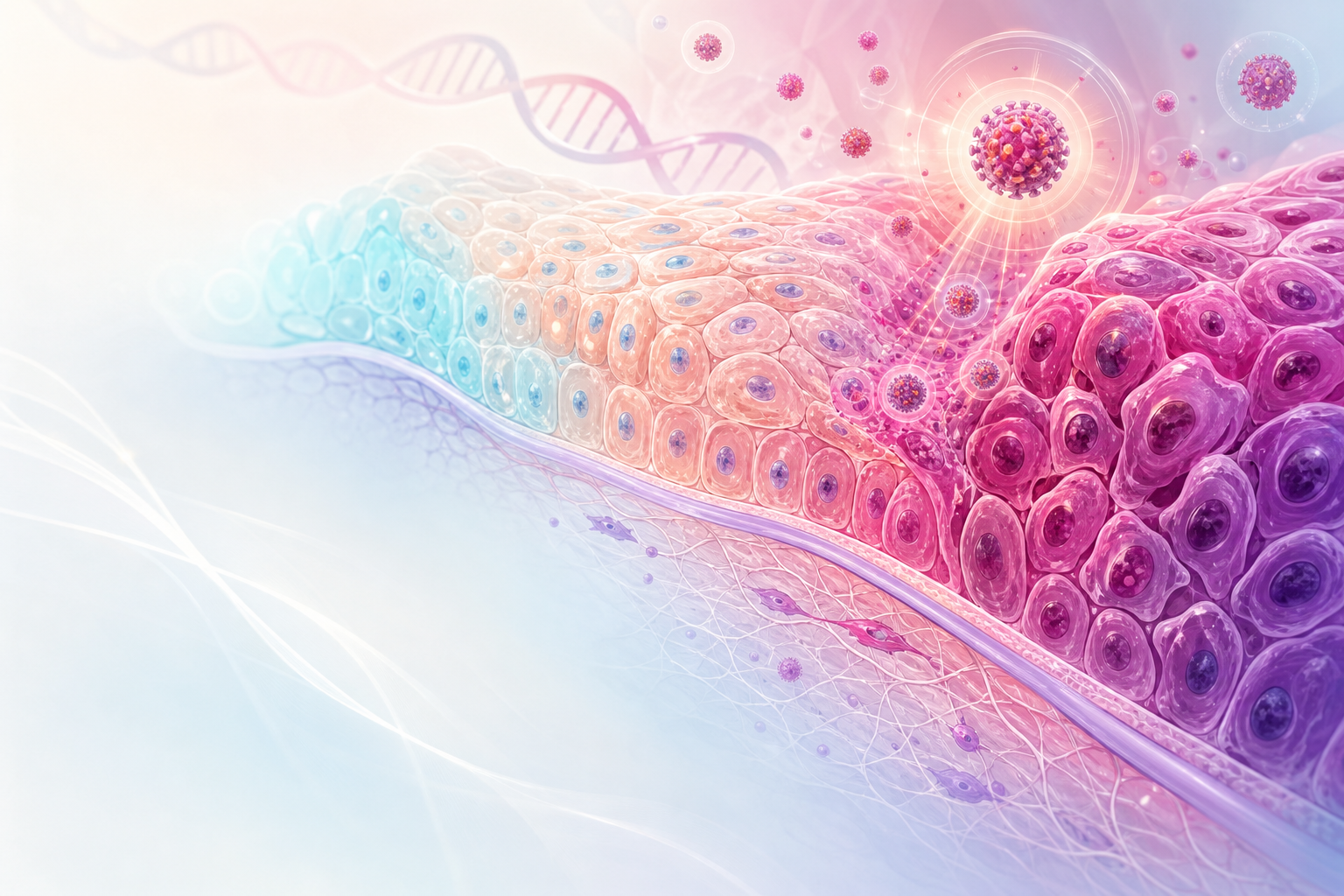
Figure D8.1 — The transformation zone and squamocolumnar junction: immature squamous metaplasia is the HPV-vulnerable field where CIN arises.
CIN does not arise randomly across the cervix; it arises almost exclusively in the transformation zone (TZ). The native ectocervix is covered by mature stratified squamous epithelium; the endocervical canal by a single layer of columnar (glandular) epithelium. The junction between them — the squamocolumnar junction (SCJ) — is not fixed. Under the influence of oestrogen, low vaginal pH (menarche, pregnancy, combined hormonal contraception), the original columnar epithelium that has everted onto the ectocervix undergoes squamous metaplasia, converting to squamous epithelium and shifting the functional SCJ inward over a woman's reproductive life.
This metaplastic transformation zone is the vulnerable site for two reasons. First, metaplasia proceeds via reserve cells — undifferentiated, mitotically active cells beneath the columnar layer — which are exactly the actively dividing basal-type cells HPV needs to infect and establish a persistent reservoir. Second, the SCJ harbours a discrete population of cuboidal junctional cells (an embryologically distinct residual population) that appear especially susceptible to HPV-driven transformation; almost all HPV-associated CIN3 and squamous carcinoma localise to this zone. The clinical corollary is that colposcopy is fundamentally an examination of the transformation zone, and that as the SCJ recedes into the canal with age, the TZ becomes less visible — making cytology and HPV testing (rather than visual inspection) the more reliable screen in older women. (See colposcopy and benign-cervical-pathology.)
HPV — the necessary cause
Cervical squamous neoplasia is, to a first approximation, an obligate consequence of persistent infection with high-risk (oncogenic) HPV genotypes. HPV is a small non-enveloped double-stranded DNA virus. Around 14 genotypes are oncogenic; HPV 16 and 18 account for roughly 70% of cervical cancers worldwide, with 16 the dominant driver of HSIL and squamous carcinoma and 18 over-represented in adenocarcinoma. Other high-risk types (31, 33, 45, 52, 58 and others) make up much of the remainder — the rationale for the broad coverage of the nonavalent (9-valent) vaccine endorsed in the WHO programme.
The virus reaches the basal/reserve cells through micro-abrasions in the metaplastic epithelium. There it can establish two distinct biological states, and the distinction is the conceptual spine of this whole topic:
-
Productive (permissive) infection. The viral genome persists as a low-copy episome (free circular DNA, not integrated). As the infected basal cell divides and its progeny differentiate upward, the virus co-opts the differentiation programme to complete its life cycle — amplifying its genome and assembling new virions in the upper layers. The cytopathic signature of this is the koilocyte: a superficial squamous cell with a perinuclear halo, nuclear enlargement and irregularity, reflecting the action of the viral E4 protein on the cytokeratin cytoskeleton. Productive infection corresponds morphologically to LSIL / CIN1 and is the cervix doing what most infected cervices do — supporting a transient, immunologically self-limiting infection.
-
Transforming (abortive) infection. When the normal coupling between differentiation and viral life cycle breaks down — classically (though not exclusively) associated with integration of the viral genome into the host chromosome — the virus can no longer complete productive replication but instead drives uncontrolled basal-type proliferation. This is HSIL (CIN2/CIN3), true precancer.
E6 and E7 — the molecular engine

Figure D8.2 — Episomal (productive, LSIL) vs integrated (transforming, HSIL) HPV DNA, and how integration drives high-grade disease.
The transforming phenotype is driven by deregulated, constitutive expression of two viral oncoproteins, E6 and E7. In the episomal state, the viral E2 protein represses the promoter that drives E6/E7. When integration occurs it typically disrupts the E2 open reading frame, releasing this brake — so E6 and E7 are now expressed continuously and at high level, even in the proliferating basal layer where they have no business being active. Their targets are the two master tumour-suppressor pathways:
-
E7 binds and degrades retinoblastoma protein (pRb). pRb normally restrains the cell cycle by sequestering the E2F transcription factors at the G1/S checkpoint. With pRb gone, E2F is freed and the cell is pushed unrestrained into S phase. A diagnostically crucial downstream consequence: loss of functional pRb removes the negative feedback on p16^INK4a^, which accumulates to high levels. This is why diffuse block-positive p16 immunohistochemistry is the morphological surrogate for transforming high-risk HPV infection and is used (per WHO 2020 / LAST) to resolve equivocal CIN2 lesions.
-
E6 binds p53 and (via the cellular ligase E6-AP) targets it for proteasomal degradation. p53 is the "guardian of the genome" — it arrests the cycle and triggers apoptosis in response to DNA damage. Its destruction means cells that acquire DNA damage are neither repaired nor culled. E6 additionally activates telomerase (hTERT), conferring replicative immortality.
The combined effect is a cell forced to proliferate (E7) while losing its ability to repair DNA, halt the cycle or die in response to damage (E6). Over years of persistent E6/E7 expression, the resulting genomic instability allows accumulation of secondary host mutations and aneuploidy. CIN is therefore not a single mutational event but a progressive accumulation; the higher the grade, the more the proliferating, abnormal cells occupy the full epithelial thickness, and the closer the lesion is to acquiring the changes needed for stromal invasion. This molecular sequence is developed further in cervical-carcinogenesis.
Morphology and the WHO 2020 / LAST two-tier system
Histologically, CIN is graded by how far up the epithelium the immature, atypical, mitotically active basal-type cells extend, with loss of normal maturation/stratification, nuclear enlargement, hyperchromasia, pleomorphism and abnormal (including suprabasal) mitoses:
| Old 3-tier (CIN) | Depth of atypical immature cells | WHO 2020 / LAST 2-tier | Biology |
|---|---|---|---|
| CIN1 | Lower third only; koilocytes prominent | LSIL | Productive HPV infection; mostly regresses |
| CIN2 | Up to lower two-thirds | HSIL (p16 resolves the borderline) | Equivocal — biologically heterogeneous |
| CIN3 | Full (or near-full) thickness | HSIL | True precancer; high persistence/progression |
The WHO Classification of Tumours of the Female Genital Tract, 5th edition (2020), harmonised with the LAST (Lower Anogenital Squamous Terminology, 2014) project, collapses the three-tier CIN system into LSIL and HSIL. The rationale is biological: LSIL ≈ CIN1 ≈ a productive, usually transient infection, whereas HSIL ≈ CIN2/CIN3 ≈ a transforming infection that is a genuine cancer precursor. CIN2 was the problem child — poorly reproducible between pathologists and biologically a mix of regressing LSIL-like lesions and true HSIL — so p16 immunohistochemistry is used to stratify it: block-positive (diffuse, full-thickness) p16 → HSIL; negative/patchy → treat as LSIL. The same framework distinguishes HPV-associated from the rarer HPV-independent squamous lesions elsewhere in the lower genital tract. The terms CIN1–3 remain in clinical use, so the registrar must be fluent in both vocabularies.
Natural history — the interruptible window

Figure D8.3 — The CIN spectrum and its natural history: regression, persistence and progression rates from CIN1/LSIL to CIN3/HSIL.
The quantitative natural history is examinable and underpins all management:
- The majority of incident HPV infections clear within 1–2 years through cell-mediated immunity, and most LSIL/CIN1 regresses spontaneously — the reason CIN1 is generally watched, not treated, in immunocompetent women.
- HSIL/CIN3 is the obligate precursor lesion; a substantial proportion, if untreated, progresses to invasive cancer over a typically protracted interval (commonly cited as on the order of 10–15 years), though the latency is variable and can be much shorter in the immunosuppressed. (The exact regression/progression percentages vary between studies; treat any single quoted figure cautiously.)
- Persistence of high-risk HPV — not mere acquisition — is the rate-limiting step. This is why a one-off positive HPV test means less than a persistently positive one, and why HPV-based screening with genotyping/triage outperforms single cytology.
HIV and the South African context — a steeper, faster pathway
This is where SA pathophysiology diverges sharply from textbook Western natural history, and it is high-yield. South Africa carries one of the world's largest HIV burdens, and HIV transforms HPV biology at every step:
- Higher acquisition and reduced clearance. CD4-mediated immunity is what clears HPV; HIV-related immunosuppression means HPV is acquired more readily, cleared less often, and a broader range of (often multiple concurrent) high-risk genotypes persist.
- Higher prevalence, higher grade, faster progression. Women living with HIV have markedly higher rates of HSIL and of CIN recurrence after treatment, and progression to invasion at younger ages. Lower CD4 counts correlate with higher-grade disease.
- ART helps but does not abolish the excess risk, so screening intensity — not just ART — remains essential.
These facts directly shape SA policy. The National Cervical Cancer Screening Guidelines and the SASOG / BetterGyn Clinical Guideline (2024) specify that HIV-positive women are screened at the time of HIV diagnosis and more frequently (approximately 3-yearly) thereafter, regardless of age — a deliberate departure from the historic public-sector cytology policy of screening HIV-negative women aged 30–50 (three smears, 10-yearly from age 30). South Africa is moving toward HPV-DNA primary screening (a direction informed by the SA DiaVACCS trial), with triage by genotyping (16/18), cytology or VIA, then treatment by thermal ablation or LLETZ — a screen-triage-treat model aligned to the WHO Cervical Cancer Elimination Strategy (90-70-90 by 2030). (Confirm exact intervals and the current screen-triage-treat algorithm against the live BetterGyn 2024 / NDoH PDF before quoting precise numbers in an exam answer.) Access constraints — NHLS turnaround for cytology/HPV testing, the school-based HPV vaccination programme (girls 9–14 since 2014, moving to a WHO-endorsed single-dose schedule with the nonavalent vaccine) — all bear directly on how the CIN window is exploited at population scale.
Assessment
CIN is by definition asymptomatic — it is a screen-detected, histology-confirmed diagnosis, never a clinical one. There is no symptom of CIN; abnormal bleeding or discharge should raise concern for established invasive disease, not CIN, and mandates examination rather than reassurance. The diagnostic pathway runs:
- Screen — high-risk HPV-DNA test (the direction of SA policy and the WHO high-performance test standard) or cervical cytology. A positive screen reflects infection or its cytological footprint, not a tissue diagnosis.
- Triage — HPV genotyping (16/18 carry the highest risk), reflex cytology, or VIA, to decide who needs colposcopy or immediate treatment.
- Colposcopy — application of acetic acid (acetowhite change reflects increased nuclear density/protein in dysplastic epithelium) and Lugol's iodine (dysplastic epithelium is glycogen-poor and stains mustard-yellow, not mahogany). The colposcopist assesses the transformation zone, vascular patterns (punctation, mosaicism, atypical vessels) and lesion margins, and directs a biopsy. (See colposcopy.)
- Histology — the definitive diagnosis. The biopsy is graded LSIL/HSIL (CIN1–3), with p16 immunohistochemistry used to resolve a morphologically equivocal CIN2 into LSIL or HSIL per WHO 2020 / LAST.
The conceptual point for assessment is that each step tests a different layer of the pathophysiology — the screen detects the virus/infection, colposcopy localises the transformed epithelium within the TZ, and histology confirms the depth of transformation that defines grade. SA-specific assessment realities (NHLS laboratory access, the practicality of a single-visit screen-and-treat in rural settings) are why VIA and thermal ablation retain a role alongside laboratory-based pathways.
Management
Management follows directly from the natural history and is covered in full in cin-management; the pathophysiological principles are:
- LSIL / CIN1 in an immunocompetent woman is usually observed with repeat HPV/cytology, because most regress as the infection clears. Treating every CIN1 would overtreat a self-limiting productive infection and risk cervical incompetence in young women.
- HSIL / CIN2–3 is treated, because it is a genuine precancer with meaningful progression risk. Treatment is either ablative (destroying the TZ — thermal/cryo ablation, suitable when the lesion and entire SCJ are fully visible and invasion excluded) or excisional (LLETZ / large-loop excision of the transformation zone, which both treats and provides a specimen to exclude occult invasion). Excision is preferred where the SCJ is not fully visible, where there is suspicion of invasion or glandular disease, or for recurrent/extensive lesions.
- Excision treats the whole transformation zone, not just the visible lesion, precisely because the TZ is the field at risk — this is the surgical translation of the metaplasia/SCJ biology.
- In women living with HIV, the threshold to treat is lower, recurrence is higher, optimising ART and surveillance is integral, and follow-up is intensified — a direct consequence of the impaired clearance and faster progression described above.
- Prevention upstream — HPV vaccination — prevents the initiating infection and is the population-level expression of the same pathophysiology, the "90" of vaccination in the WHO 90-70-90 strategy.
A balance against over-treatment is essential: LLETZ is associated with later preterm birth and second-trimester loss (cervical length/competence effects), so treating self-limiting CIN1 in a young woman carries real reproductive cost — a recurring exam discrimination point.
Red flags / pitfalls
- Symptoms mean invasion, not CIN. Postcoital, intermenstrual or postmenopausal bleeding, or a visible/friable cervical lesion, must be examined and biopsied for cancer — never attributed to "CIN" and never managed as a screening abnormality. CIN is silent.
- A normal-looking cervix does not exclude HSIL. Do not let a clinically unremarkable cervix override a positive screen — the lesion lives in the transformation zone and is invisible to the naked eye.
- The SCJ recedes with age. In older and postmenopausal women the transformation zone is endocervical and may be invisible at colposcopy (type 3 TZ) — visual methods (VIA) lose sensitivity and excision rather than ablation may be needed to sample the canal.
- Don't over-call or under-treat CIN2. It is the least reproducible grade; use p16 to stratify it into LSIL or HSIL rather than defaulting to treatment of every CIN2 in a young woman.
- HIV status changes everything. Treating CIN in a woman living with HIV by an immunocompetent algorithm under-screens and under-treats her — she needs earlier, more frequent screening and intensified follow-up.
- HPV positivity ≠ disease. A single positive HPV test reflects infection, much of which clears; it is persistence and genotype/triage that determine risk. Conversely, do not falsely reassure: persistent HPV with HSIL on histology is precancer.
- Glandular and HPV-independent lesions hide. This chapter is squamous CIN; adenocarcinoma-in-situ and HPV-independent lesions behave differently, are harder to detect colposcopically, and warrant excision/specialist referral.
Evidence anchors
- WHO Classification of Tumours of the Female Genital Tract, 5th edition (2020) — the histological typing and the LAST/2014-harmonised LSIL/HSIL two-tier terminology (replacing CIN1–3), HPV-associated vs HPV-independent squamous lesions, and the role of p16 immunohistochemistry in resolving CIN2.
- South African cervical cancer screening — SASOG / BetterGyn Clinical Guideline (2024), with the NDoH National Cervical Cancer Screening / Prevention & Control Policy and National Cervical Cancer Screening Guidelines — SA screen-triage-treat model, the move toward HPV-DNA primary screening (informed by the DiaVACCS trial), the historic cytology policy (women 30–50, 3 smears 10-yearly from 30), and intensified screening for HIV-positive women (at diagnosis and ~3-yearly regardless of age).
- WHO Cervical Cancer Elimination Strategy (2020) — 90-70-90 by 2030 — vaccination, high-performance screening, and treatment targets; single-dose HPV schedule (WHO 2022) and nonavalent vaccine coverage underpinning SA's school-based programme.
- ESGO/ESTRO/ESP Guidelines for cervical cancer — Update 2023 — for the precancer-to-cancer continuum, FIGO 2018 staging context for what CIN can become, and pathology principles.
