Clinical overview
Melanoma of the female genital tract is rare but disproportionately lethal, and it is a tumour the FCOG(SA) registrar must be able to recognise pathologically because the diagnosis is so easily missed clinically. Mucosal melanoma of the vulva and vagina accounts for roughly 5–10% of all vulval malignancies and a smaller fraction of vaginal cancers, yet it is the second commonest vulval malignancy after squamous cell carcinoma. The vulva is the single most frequent site of female genital melanoma, with the labia minora, clitoris and the mucocutaneous junction of the introitus being characteristic locations; the vagina (most often the lower third and anterior wall) and, rarely, the cervix and urethra follow. The peak incidence is in postmenopausal women in the sixth to eighth decades, although it occurs at any age.
What makes this tumour dangerous is biology, not just rarity. Genital mucosal melanoma is fundamentally a different disease from cutaneous melanoma: it is not driven by ultraviolet radiation, it carries a lower mutational burden, a distinct molecular profile, and a markedly worse stage-for-stage prognosis — five-year survival figures of roughly 10–25% for vaginal and 25–50% for vulval primaries reflect both intrinsically aggressive behaviour and late presentation. Pigmented vulval lesions are frequently dismissed as benign naevi, seborrhoeic keratoses, angiokeratomas or simply attributed to physiological hyperpigmentation, and amelanotic melanomas masquerade as granulation tissue, polyps or even a Bartholin's cyst. For the South African registrar working in a high-HIV-prevalence, resource-variable setting, the practical message is that any new, changing, pigmented, ulcerated or bleeding vulvovaginal lesion warrants a biopsy with melanoma explicitly on the differential — this is a tumour where pattern recognition and a low threshold to biopsy save lives. This objective concerns the pathological features, so the weight below sits in Core knowledge: gross morphology, microscopic architecture and cytology, the immunohistochemical panel, and the molecular landscape that increasingly drives systemic therapy.
Core knowledge
Origin and the WHO framework
Melanocytes are neural-crest-derived dendritic cells that normally reside scattered along the basal layer of squamous epithelium. In the vulva they populate keratinised skin, the mucocutaneous transition zone and glabrous mucosa; in the vagina and cervix they are present in a minority of women as an embryological remnant, which is why primary vaginal and cervical melanomas can arise at all. Melanoma is the malignant proliferation of these melanocytes. The authoritative typing framework is the WHO Classification of Tumours of the Female Genital Tract, 5th edition (2020), which classifies genital melanocytic lesions and recognises the distinction between mucosal (mucocutaneous) melanoma and rarer purely cutaneous vulval melanoma, alongside the benign melanocytic differential (lentigo, melanosis, genital naevi including the histologically alarming atypical melanocytic naevus of the genital type, "AMNGT", which must not be over-diagnosed as melanoma).
Gross / macroscopic features
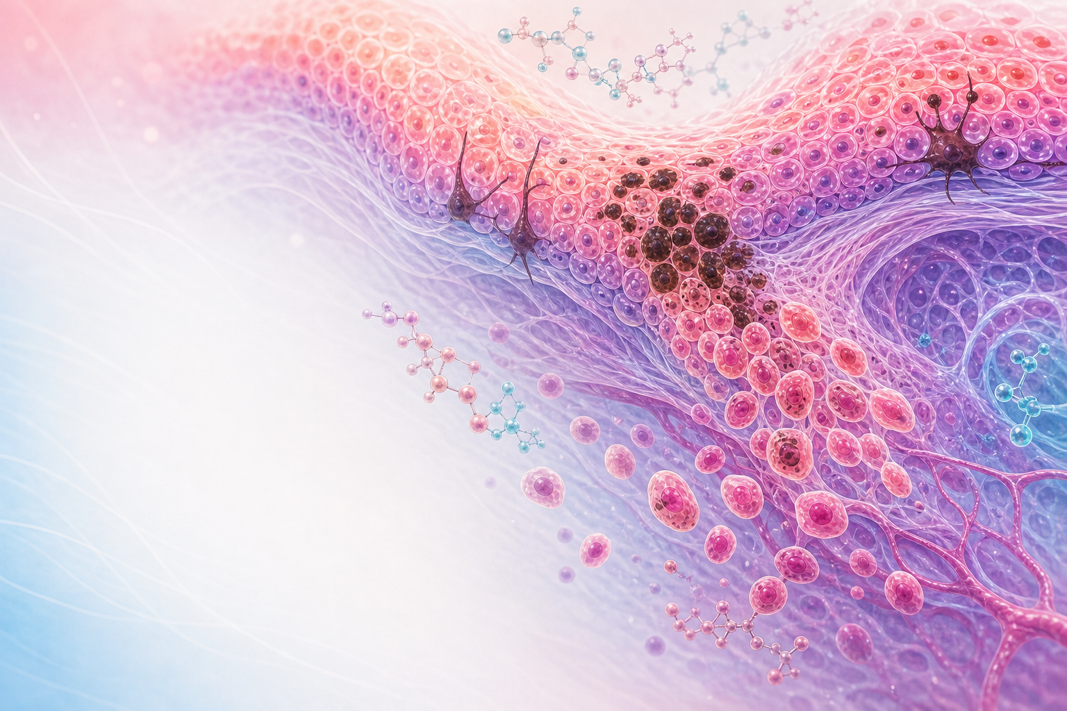
Figure D16.1 — Mucosal melanoma, easy to miss: arises from mucosal melanocytes, often amelanotic, the ABCDE warning features, and Breslow-thickness staging.
Macroscopic recognition is the first diagnostic gate, and it follows the same logic as the ABCDE rule adapted to mucosa:
- Asymmetry, irregular Borders, Colour variegation — true melanomas are typically asymmetrical with notched, ill-defined margins and a mottled mixture of black, brown, blue-grey, tan, pink and depigmented (white/regressed) zones. Uniform, symmetrical, evenly pigmented small lesions are more reassuring but never exempt from biopsy if changing.
- Diameter and Evolution — lesions are often >7 mm and, crucially, have changed (enlarged, darkened, become raised, ulcerated or begun to bleed).
- Pigmentation is variable: most are pigmented (brown-black macule, plaque, polypoid or nodular mass), but a clinically critical minority — perhaps a quarter of vulvovaginal melanomas — are amelanotic or hypomelanotic, appearing as flesh-coloured, pink, red or ulcerated nodules. Amelanotic melanoma is a notorious diagnostic trap and a frequent cause of delay.
- Surface and growth pattern: ranges from a flat spreading macule to an exophytic, polypoid or fungating ulcerated mass. Ulceration and bleeding are adverse features and correlate with advanced primary tumour thickness.
- Satellite lesions and multifocality can be present, reflecting radial intra-epithelial spread or in-transit metastasis.
Microscopic features — architecture
Histology is the diagnostic gold standard. Two architectural components are assessed: the intra-epithelial (in-situ / radial-growth) component and the dermal/stromal invasive (vertical-growth) component.
- Junctional and intra-epithelial spread: malignant melanocytes proliferate along the dermo-epidermal (or epithelial–stromal) junction as single atypical cells and irregular, confluent nests of varying size and shape, with upward (pagetoid) migration of single melanocytes into the upper epithelial layers. Pagetoid spread is an important clue and forms part of the differential with extramammary Paget disease and pagetoid HSIL.
- Lentiginous / mucosal-lentiginous pattern: the commonest growth pattern of mucosal genital melanoma is a mucosal-lentiginous melanoma — a confluent basal proliferation of atypical melanocytes along an often-elongated rete pattern, analogous to acral-lentiginous melanoma. Superficial-spreading and nodular patterns also occur, the nodular type having little or no radial phase and proceeding rapidly to vertical (invasive) growth.
- Invasion and vertical growth: invasive melanoma extends as sheets, nests or single cells into the underlying stroma, frequently with an expansile dermal nodule. Lymphovascular and perineural invasion may be seen and are adverse.
- Regression (a zone of fibrosis, melanophages and lymphocytes where tumour has involuted) and tumour-infiltrating lymphocytes (TILs) are documented; brisk TILs carry prognostic weight in cutaneous disease and are noted in mucosal disease, though their prognostic strength is less firmly established here.
Microscopic features — cytology
The malignant melanocyte is morphologically protean — melanoma is the classic histological "great imitator". Recognisable cytological clues include:
- Large epithelioid cells with abundant eosinophilic-to-amphophilic cytoplasm, large nuclei, prominent (often single, cherry-red) macronucleoli, and intranuclear cytoplasmic pseudo-inclusions.
- Spindle-cell morphology, which can closely mimic a sarcoma, smooth-muscle tumour or spindle-cell SCC; spindled and desmoplastic variants are particularly treacherous and may be amelanotic.
- Variable melanin pigment within tumour cells and in stromal melanophages; pigment may be abundant, focal or absent.
- Frequent and atypical mitoses — mitotic activity (mitoses per mm²) is a recognised prognostic determinant. Other variants documented at genital sites include small-cell, balloon-cell, signet-ring and clear-cell forms, reinforcing why melanoma must be on the differential for almost any poorly differentiated genital malignancy.
Microscopic staging of the primary — thickness and depth
Because melanoma prognosis is so tightly bound to how deep the tumour has invaded, the pathology report must quantify it:
- Breslow thickness — the vertical depth in millimetres from the top of the granular layer (or the base of an ulcer) to the deepest tumour cell — is the single most important prognostic histological parameter and the basis of the tumour (T) category. It applies well to keratinised vulval skin; on glabrous mucosa lacking a granular layer it is measured from the surface, which the reporting pathologist should note.
- Clark level (anatomical level of invasion) is of historical and adjunctive value but is superseded by Breslow thickness.
- Ulceration status and mitotic rate are reported as independent adverse prognostic features.
- The applicability of cutaneous thickness cut-offs to mucosal disease is imperfect, which is one reason mucosal genital melanoma is frequently staged using both melanoma-specific (AJCC/TNM) and FIGO anatomical frameworks; some centres apply the FIGO system used for the corresponding squamous-cell site (e.g. FIGO 2018 for cervical primaries, or the vulval/vaginal FIGO schemes) as an anatomical adjunct. The registrar should state thickness, ulceration, mitoses, margins, lymphovascular invasion and node status, and recognise that no staging system performs as well for mucosal melanoma as for cutaneous disease — flag this honestly rather than forcing the tumour into a cutaneous box.
Immunohistochemistry — confirming the diagnosis
When melanin is scant or the morphology is spindled/epithelioid and undifferentiated, immunohistochemistry confirms melanocytic lineage and is essential to separate melanoma from its mimics (SCC, sarcoma, lymphoma, extramammary Paget disease, neuroendocrine tumours):
- S100 — highly sensitive, stains nuclei and cytoplasm, positive in nearly all melanomas including spindled/desmoplastic variants; not specific (also positive in nerve-sheath tumours, Langerhans cells, some carcinomas).
- SOX10 — sensitive nuclear marker, useful and reliable including in spindled/desmoplastic melanoma.
- HMB-45 — specific but less sensitive; marks gp100, often patchy or negative in spindle/desmoplastic melanoma.
- Melan-A / MART-1 and tyrosinase — melanocyte-differentiation markers, specific, variably sensitive (less so in spindled tumours).
- PRAME — increasingly used to support malignancy and help distinguish melanoma from benign naevi (including the genital atypical naevus).
- Ki-67 / proliferation index supports malignancy.
A practical panel pairs a sensitive marker (S100 or SOX10) with specific markers (HMB-45, Melan-A). A useful pitfall to remember: melanoma can aberrantly express cytokeratins, so a "keratin-positive" undifferentiated genital tumour is not automatically a carcinoma — always confirm with melanocytic markers when morphology is ambiguous.
Molecular pathology
The molecular profile of mucosal genital melanoma differs sharply from sun-exposed cutaneous melanoma and is clinically actionable:
- BRAF V600 mutations — common in cutaneous melanoma but uncommon in mucosal/vulvovaginal melanoma (a minority only). This matters because BRAF status is tested to determine eligibility for BRAF/MEK-targeted therapy; most genital mucosal melanomas are BRAF wild-type.
- KIT (c-KIT) mutations/amplifications — enriched in mucosal and acral melanoma relative to cutaneous disease, and a potential target for KIT inhibitors. KIT testing is therefore prioritised in genital primaries.
- NRAS mutations occur in a subset.
- Mucosal melanomas carry a lower total mutational/UV-signature burden and show more structural/copy-number changes, which is part of why response to immune checkpoint inhibition, while real, is generally less robust than in high-mutational-burden cutaneous melanoma.
A note on systemic-therapy evidence and verified sources. The molecular and immunotherapy principles above are correct in direction, but the named genital-oncology trials that anchor other Domain D chapters — pembrolizumab (KEYNOTE-A18/826), dostarlimab (RUBY), pembrolizumab–lenvatinib (KEYNOTE-775), and the ESGO/ESTRO/ESP and FIGO 2018/2023 staging frameworks — are cervical and endometrial evidence, not melanoma trials, and the project's
docs/VERIFIED-SOURCES.mddoes not list a melanoma-specific checkpoint-inhibitor or BRAF/MEK trial. I have therefore not cited any specific melanoma drug trial, dose or regimen by name; treat systemic-therapy specifics for genital melanoma as outside the verified-source set and confirm against a current melanoma guideline (e.g. ESMO/NCCN melanoma) before quoting agents or doses in an exam answer.
Assessment

Figure D16.2 — Why mucosal melanoma is so aggressive: early lymphatic and haematogenous spread, poor prognosis, and the principles of management.
Diagnosis rests on a high index of suspicion and histology. Take a focused history of the lesion's onset, change, bleeding, itch or pain, prior pigmented lesions, sun-unrelated risk factors, immunosuppression (including HIV status, given the South African epidemiology) and family history. Examine the entire lower genital tract and skin, document size, site, colour, ulceration, multifocality and inguinal nodes, and use a measuring diagram or photograph.
The decisive investigation is biopsy. For a small lesion an excisional biopsy with a narrow margin is preferred so the pathologist receives the full thickness and can measure Breslow depth; for large or multifocal lesions a generous full-thickness incisional/punch biopsy of the thickest, most atypical (and any ulcerated) area is acceptable — superficial shave biopsy is discouraged because it truncates depth measurement. The histology request should explicitly raise melanoma so the laboratory applies the correct immunohistochemical panel. Insist the report states: histological type and growth pattern, Breslow thickness, ulceration, mitotic rate, lymphovascular and perineural invasion, margin status and the immuno/molecular results (S100/SOX10, HMB-45, Melan-A, and BRAF and KIT where systemic therapy is contemplated). In the SA setting these analyses are performed through the NHLS anatomical-pathology service; molecular testing access varies by region and may require referral to a tertiary or academic laboratory — name this access gap rather than assume universal availability.
Staging work-up assesses regional and distant spread: clinical and radiological inguinofemoral/pelvic node evaluation, and cross-sectional imaging (CT chest/abdomen/pelvis; MRI for local extent of vaginal/vulval primaries; PET-CT and brain imaging where available and indicated for higher-stage disease). Sentinel lymph node biopsy has an established role in selected vulval primaries; note that this is best validated for vulval squamous cancer in the ESGO vulvar cancer guideline (2023 update), and its routine value in vulvar melanoma is less firmly established — extrapolate with caution.
Management
Management is principle-level for a pathology objective, and is multidisciplinary, centred on surgery for localised disease.
- Surgery is the mainstay for resectable primaries: wide local excision with the widest clear margins anatomy permits, prioritising negative margins over the radical, mutilating vulvectomies of the past, since extensive surgery has not been shown to improve survival in mucosal melanoma. Vaginal primaries are surgically challenging and frequently require radiotherapy.
- Regional nodes: sentinel node assessment/lymphadenectomy in selected vulval cases as above; clinically involved nodes are addressed surgically and/or with radiotherapy.
- Radiotherapy is used adjuvantly for local control (mucosal melanoma is relatively radioresistant but RT improves local control), and palliatively.
- Systemic therapy: the modern landscape for advanced/metastatic melanoma is immune checkpoint inhibition and, for the minority that are BRAF V600-mutant, BRAF/MEK-targeted therapy, with KIT inhibitors explored for KIT-altered mucosal disease — but, as flagged above, the specific agents, regimens and doses are not in this project's verified-source set, so confirm against a current dedicated melanoma guideline before quoting them. Access to checkpoint inhibitors and targeted agents is constrained in the South African public sector and is not a routine EML item for this indication at the time of writing — manage expectations and refer to a medical-oncology multidisciplinary meeting.
- Prognosis is guarded and dominated by Breslow thickness, ulceration, nodal status and distant spread; counsel honestly. See also vulval-carcinoma and vaginal-tumours for the squamous neighbours these tumours are confused with.
Red flags / pitfalls
- Amelanotic melanoma — a pink/red/ulcerated nodule with no pigment is melanoma until immunohistochemistry says otherwise; this is the commonest reason for missed diagnosis.
- Calling it "just a naevus" — never assume; the benign atypical melanocytic naevus of the genital type is histologically alarming and is itself frequently over-diagnosed as melanoma, so an experienced gynaecological/dermatopathologist (via the NHLS and referral if needed) should review difficult pigmented lesions. Both errors — false-negative and false-positive — are costly.
- Shave biopsy of a suspected melanoma destroys the ability to measure Breslow thickness and understages the tumour; take a full-thickness sample.
- "Keratin-positive, therefore carcinoma" — melanoma can express cytokeratins; always include melanocytic markers (S100/SOX10) when an undifferentiated genital tumour is being worked up.
- Spindle-cell and desmoplastic melanoma mimic sarcoma and may be S100-positive but HMB-45/Melan-A-negative — use SOX10 and keep melanoma on the differential of any spindle-cell genital lesion.
- Assuming BRAF positivity — most genital mucosal melanomas are BRAF wild-type, so do not promise BRAF-targeted therapy before testing; prioritise KIT testing in mucosal disease.
- Delayed referral — vulvovaginal melanoma is aggressive and stage-driven; do not "watch" a changing pigmented lesion. Biopsy and refer to a gynaecological-oncology MDT.
- Forcing a cutaneous stage onto a mucosal tumour — staging systems underperform here; report the raw pathology (thickness, ulceration, mitoses, margins, nodes) so the MDT can stage appropriately.
Evidence anchors
- WHO Classification of Tumours of the Female Genital Tract, 5th edition (2020) — the authoritative histological typing framework for genital melanocytic lesions and the benign differential (genital naevi, melanosis), and the source of the LSIL/HSIL (with CIN1–3 equivalence) terminology used for the squamous neighbours melanoma is confused with.
- ESGO Guidelines for vulvar cancer — Update 2023 (Int J Gynecol Cancer) — surgical-margin and sentinel-lymph-node principles for vulvar malignancy; note these are validated primarily for vulvar squamous carcinoma and are extrapolated to vulvar melanoma with caution.
- Anatomical staging context — FIGO frameworks used for the corresponding squamous sites (e.g. FIGO 2018 for cervical primaries) may be applied as an anatomical adjunct, but no staging system performs for mucosal melanoma as it does for cutaneous disease (flagged as a recognised limitation, not a melanoma-specific FIGO scheme).
- South African service context — histopathology and immunohistochemistry via NHLS anatomical pathology; molecular (BRAF/KIT) testing access varies by region and may require academic-centre referral; checkpoint-inhibitor and targeted systemic therapy for melanoma are not routine SA public-sector/EML provisions for this indication — confirm local availability.
- Uncertainty flagged honestly:
docs/VERIFIED-SOURCES.mdcontains no melanoma-specific trial, drug, dose or staging citation. The cervical/endometrial immunotherapy trials named elsewhere in Domain D (KEYNOTE-A18/826, RUBY/dostarlimab, KEYNOTE-775 pembrolizumab–lenvatinib) and the molecular-staging frameworks (FIGO 2023 endometrial; ESGO/ESTRO/ESP cervical 2023, endometrial 2025) do not apply to melanoma and are deliberately not cited as melanoma evidence. For systemic-therapy specifics, verify against a current dedicated melanoma guideline (e.g. ESMO/NCCN melanoma) before asserting agents or doses.
